 Movie
Movie Controller


+ Open data

- Basic information
| Entry | Database: PDB / ID: 3fwu | ||||||
|---|---|---|---|---|---|---|---|
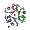
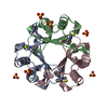
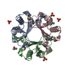
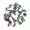
| Title | Crystal structure of Leishmania major MIF1 | ||||||
 Components Components | Macrophage migration inhibitory factor-like protein | ||||||
 Keywords Keywords | CYTOKINE / homotrimer / tautomerase | ||||||
| Function / homology |  Function and homology information Function and homology informationphenylpyruvate tautomerase / L-dopachrome isomerase / dopachrome isomerase activity / phenylpyruvate tautomerase activity / cytokine activity / : Similarity search - Function | ||||||
| Biological species |  Leishmania major (eukaryote) Leishmania major (eukaryote) | ||||||
| Method |  X-RAY DIFFRACTION / SYNCHROTRON / MOLECULAR REPLACEMENT / molecular replacement / Resolution: 1.8 Å X-RAY DIFFRACTION / SYNCHROTRON / MOLECULAR REPLACEMENT / molecular replacement / Resolution: 1.8 Å | ||||||
 Authors Authors | RIchardson, J.M. / Walkinshaw, M.D. | ||||||
 Citation Citation | Journal: Biochem.Biophys.Res.Commun. / Year: 2009 Title: Structures of Leishmania major orthologues of macrophage migration inhibitory factor Authors: Richardson, J.M. / Morrison, L.S. / Bland, N.D. / Bruce, S. / Coombs, G.H. / Mottram, J.C. / Walkinshaw, M.D. | ||||||
| History |
|
- Structure visualization
| Structure viewer | Molecule: MolmilJmol/JSmol |
|---|
- Downloads & links
-Download
| PDBx/mmCIF format | 3fwu.cif.gz | 36.5 KB | Display | PDBx/mmCIF format |
|---|---|---|---|---|
| PDB format | pdb3fwu.ent.gz | 23.9 KB | Display | PDB format |
| PDBx/mmJSON format | 3fwu.json.gz | Tree view | PDBx/mmJSON format | |
| Others |  Other downloads Other downloads |
-Validation report
| Arichive directory | https://data.pdbj.org/pub/pdb/validation_reports/fw/3fwuftp://data.pdbj.org/pub/pdb/validation_reports/fw/3fwu | HTTPS FTP |
|---|
-Related structure data
| Related structure data |  3fwtC  1uizS C: citing same article ( S: Starting model for refinement |
|---|---|
| Similar structure data |


-Links
 PDBj
PDBj- Assembly
| Deposited unit | 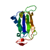
| ||||||||
|---|---|---|---|---|---|---|---|---|---|
| 1 | 
| ||||||||
| Unit cell |
|
-Components
| #1: Protein | Mass: 14697.716 Da / Num. of mol.: 1 Source method: isolated from a genetically manipulated source Source: (gene. exp.) Leishmania major (eukaryote) / Strain: Friedlin / Gene: LmjF33.1740 / Plasmid: pET28a / Production host:  |
|---|---|
| #2: Water | ChemComp-HOH /  Mass: 18.015 Da / Num. of mol.: 56 / Source method: isolated from a natural source / Formula: H2O Mass: 18.015 Da / Num. of mol.: 56 / Source method: isolated from a natural source / Formula: H2O |
-Experimental details
-Experiment
| Experiment | Method: X-RAY DIFFRACTION / Number of used crystals: 1 |
|---|
- Sample preparation
| Crystal | Density Matthews: 1.76 Å3/Da / Density % sol: 29.93 % |
|---|---|
| Crystal grow | Temperature: 290 K / Method: vapor diffusion, hanging drop / pH: 6.5 Details: 30% w/v PEG 4000, 100mM Imidazole, pH 6.5, Hanging drop, temperature 290K, VAPOR DIFFUSION, HANGING DROP |
-Data collection
| Diffraction | Mean temperature: 77 K |
|---|---|
| Diffraction source | Source: SYNCHROTRON / Site: ESRF  / Beamline: BM14 / Wavelength: 0.9765 Å / Beamline: BM14 / Wavelength: 0.9765 Å |
| Detector | Type: MAR CCD 165 mm / Detector: CCD / Date: Jun 6, 2006 |
| Radiation | Protocol: SINGLE WAVELENGTH / Monochromatic (M) / Laue (L): M / Scattering type: x-ray |
| Radiation wavelength | Wavelength: 0.9765 Å / Relative weight: 1 |
| Reflection | Resolution: 1.8→25 Å / Num. obs: 9160 / % possible obs: 98.8 % / Redundancy: 5.7 % / Biso Wilson estimate: 18.3 Å2 / Rmerge(I) obs: 0.069 |
| Reflection shell | Resolution: 1.8→1.9 Å / Redundancy: 3.5 % / Rmerge(I) obs: 0.121 / Mean I/σ(I) obs: 7.2 / Num. unique all: 1242 / % possible all: 93.4 |
-Phasing
| Phasing | Method: molecular replacement |
|---|
- Processing
| Software |
| |||||||||||||||||||||||||||||||||||||||||||||||||||||||||||||||||
|---|---|---|---|---|---|---|---|---|---|---|---|---|---|---|---|---|---|---|---|---|---|---|---|---|---|---|---|---|---|---|---|---|---|---|---|---|---|---|---|---|---|---|---|---|---|---|---|---|---|---|---|---|---|---|---|---|---|---|---|---|---|---|---|---|---|---|
| Refinement | Method to determine structure: MOLECULAR REPLACEMENT Starting model: PDB entry 1UIZ Resolution: 1.8→21.54 Å / Cor.coef. Fo:Fc: 0.943 / Cor.coef. Fo:Fc free: 0.88 / Occupancy max: 1 / Occupancy min: 0.5 / SU B: 3.85 / SU ML: 0.118 / Cross valid method: THROUGHOUT / σ(F): 0 / ESU R: 0.161 / ESU R Free: 0.16 / Stereochemistry target values: MAXIMUM LIKELIHOOD Details: HYDROGENS HAVE BEEN ADDED IN THE RIDING POSITIONS, U VALUES REFINED INDIVIDUALLY
| |||||||||||||||||||||||||||||||||||||||||||||||||||||||||||||||||
| Solvent computation | Ion probe radii: 0.8 Å / Shrinkage radii: 0.8 Å / VDW probe radii: 1.4 Å / Solvent model: MASK | |||||||||||||||||||||||||||||||||||||||||||||||||||||||||||||||||
| Displacement parameters | Biso max: 49.23 Å2 / Biso mean: 18.299 Å2 / Biso min: 9.41 Å2
| |||||||||||||||||||||||||||||||||||||||||||||||||||||||||||||||||
| Refinement step | Cycle: LAST / Resolution: 1.8→21.54 Å
| |||||||||||||||||||||||||||||||||||||||||||||||||||||||||||||||||
| Refine LS restraints |
| |||||||||||||||||||||||||||||||||||||||||||||||||||||||||||||||||
| LS refinement shell | Resolution: 1.8→1.847 Å / Total num. of bins used: 20
|
