[English] 日本語
Yorodumi
- PDB-6fvh: Macrophage Migration Inhibitory Factor (MIF) with Covalently Boun... -

+
Open data


ID or keywords:

Loading...

-
Basic information

Entry
Database: PDB / ID: 6fvh
Title
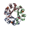
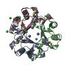
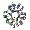
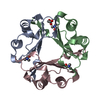
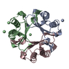
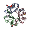
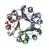
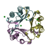
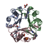
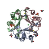
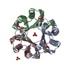
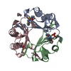
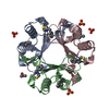
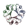
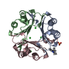
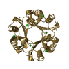
Macrophage Migration Inhibitory Factor (MIF) with Covalently Bound PITC
ComponentsMacrophage migration inhibitory factor
KeywordsISOMERASE / ISOMERASE INHIBITOR
Function / homology
Function and homology information


positive regulation of prostaglandin secretion involved in immune response / positive regulation of myeloid leukocyte cytokine production involved in immune response / dopachrome isomerase activity / phenylpyruvate tautomerase / L-dopachrome isomerase / phenylpyruvate tautomerase activity / cytokine receptor binding / positive regulation of lipopolysaccharide-mediated signaling pathway / positive regulation of arachidonic acid secretion / negative regulation of myeloid cell apoptotic process ...positive regulation of prostaglandin secretion involved in immune response / positive regulation of myeloid leukocyte cytokine production involved in immune response / dopachrome isomerase activity / phenylpyruvate tautomerase / L-dopachrome isomerase / phenylpyruvate tautomerase activity / cytokine receptor binding / positive regulation of lipopolysaccharide-mediated signaling pathway / positive regulation of arachidonic acid secretion / negative regulation of myeloid cell apoptotic process / negative regulation of macrophage chemotaxis / negative regulation of mature B cell apoptotic process / positive regulation of chemokine (C-X-C motif) ligand 2 production / negative regulation of protein metabolic process / prostaglandin biosynthetic process / carboxylic acid metabolic process / regulation of macrophage activation / negative regulation of intrinsic apoptotic signaling pathway in response to DNA damage by p53 class mediator / chemoattractant activity / protein homotrimerization / positive regulation of protein kinase A signaling / negative regulation of cellular senescence / negative regulation of DNA damage response, signal transduction by p53 class mediator / DNA damage response, signal transduction by p53 class mediator / positive regulation of B cell proliferation / positive regulation of phosphorylation / Gene and protein expression by JAK-STAT signaling after Interleukin-12 stimulation / negative regulation of cell migration / cytokine activity / positive regulation of cytokine production / Cell surface interactions at the vascular wall / positive regulation of fibroblast proliferation / positive regulation of peptidyl-tyrosine phosphorylation / cellular senescence / positive regulation of tumor necrosis factor production / positive regulation of peptidyl-serine phosphorylation / secretory granule lumen / protease binding / vesicle / ficolin-1-rich granule lumen / positive regulation of ERK1 and ERK2 cascade / cell surface receptor signaling pathway / inflammatory response / negative regulation of gene expression / innate immune response / positive regulation of cell population proliferation / Neutrophil degranulation / negative regulation of apoptotic process / cell surface / extracellular space / extracellular exosome / extracellular region / nucleoplasm / identical protein binding / plasma membrane / cytosol / cytoplasm
Similarity search - Function
Macrophage migration inhibitory factor, conserved site / Macrophage migration inhibitory factor family signature. / Macrophage migration inhibitory factor / Macrophage migration inhibitory factor (MIF) / Macrophage Migration Inhibitory Factor / Macrophage Migration Inhibitory Factor / Tautomerase/MIF superfamily / 2-Layer Sandwich / Alpha Beta
Similarity search - Domain/homology
N-phenylthioformamide / Macrophage migration inhibitory factor
Similarity search - Component
Biological speciesHomo sapiens (human)
MethodX-RAY DIFFRACTION / SYNCHROTRON / MOLECULAR REPLACEMENT / Resolution: 1.4 Å
AuthorsSamygina, V.R. / Bourenkov, G. / Sokolov, A.V.
Funding support Russian Federation, 1items
OrganizationGrant numberCountry
RFBR15-54-74006 Russian Federation
CitationJournal: Biochemistry Mosc. / Year: 2018
Title: Structural Study of the Complex Formed by Ceruloplasmin and Macrophage Migration Inhibitory Factor.
Authors: Sokolov, A.V. / Dadinova, L.A. / Petoukhov, M.V. / Bourenkov, G. / Dubova, K.M. / Amarantov, S.V. / Volkov, V.V. / Kostevich, V.A. / Gorbunov, N.P. / Grudinina, N.A. / Vasilyev, V.B. / Samygina, V.R.
History
DepositionMar 2, 2018Deposition site: PDBE / Processing site: PDBE
Revision 1.0Jun 6, 2018Provider: repository / Type: Initial release
Revision 1.1Sep 19, 2018Group: Data collection / Database references / Category: citation
Item: _citation.country / _citation.journal_abbrev ..._citation.country / _citation.journal_abbrev / _citation.journal_id_CSD / _citation.journal_id_ISSN / _citation.journal_volume / _citation.page_first / _citation.page_last / _citation.pdbx_database_id_DOI / _citation.pdbx_database_id_PubMed / _citation.title / _citation.year
Revision 1.2Jan 17, 2024Group: Data collection / Database references / Refinement description
Category: chem_comp_atom / chem_comp_bond ...chem_comp_atom / chem_comp_bond / database_2 / pdbx_initial_refinement_model
Item: _database_2.pdbx_DOI / _database_2.pdbx_database_accession

-
Structure visualization

Structure viewerMolecule:
MolmilJmol/JSmol

Downloads & links

-
Assembly

Deposited unit
A: Macrophage migration inhibitory factor
B: Macrophage migration inhibitory factor
C: Macrophage migration inhibitory factor
hetero molecules


Theoretical massNumber of molelcules
Total (without water)38,05312
Polymers37,0653
Non-polymers9889
Water8,125451
1


  • Idetical with deposited unit
  • defined by author&software
  • Evidence: gel filtration
TypeNameSymmetry operationNumber
identity operation1_555x,y,z1
Buried area7450 Å2
ΔGint-90 kcal/mol
Surface area13650 Å2
MethodPISA
Unit cell
Length a, b, c (Å)67.822, 68.375, 88.596
Angle α, β, γ (deg.)90.00, 90.00, 90.00
Int Tables number19
Space group name H-MP212121

-
Components

#1: Protein Macrophage migration inhibitory factor / / MIF / Glycosylation-inhibiting factor / GIF / L-dopachrome isomerase / L-dopachrome tautomerase / ...MIF / Glycosylation-inhibiting factor / GIF / L-dopachrome isomerase / L-dopachrome tautomerase / Phenylpyruvate tautomerase


Mass: 12355.056 Da / Num. of mol.: 3
Source method: isolated from a genetically manipulated source
Source: (gene. exp.) Homo sapiens (human) / Gene: MIF, GLIF, MMIF / Production host: Escherichia coli (E. coli)
References: UniProt: P14174, phenylpyruvate tautomerase, L-dopachrome isomerase
#2: Chemical
ChemComp-SO4 / SULFATE ION / Sulfate


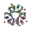
Mass: 96.063 Da / Num. of mol.: 6 / Source method: obtained synthetically / Formula: SO4
#3: Chemical ChemComp-0FI / N-phenylthioformamide / PHENYL ISOTHIOCYANATE, bound form / Phenyl isothiocyanate


Mass: 137.202 Da / Num. of mol.: 3 / Source method: obtained synthetically / Formula: C7H7NS
#4: Water ChemComp-HOH / water / Water


Mass: 18.015 Da / Num. of mol.: 451 / Source method: isolated from a natural source / Formula: H2O

-
Experimental details

-
Experiment

ExperimentMethod: X-RAY DIFFRACTION / Number of used crystals: 1

-
Sample preparation

CrystalDensity Matthews: 2.75 Å3/Da / Density % sol: 55.27 %
Crystal growTemperature: 293 K / Method: vapor diffusion, hanging drop / pH: 7 / Details: Ammonium Sulfate, Hepes / PH range: 7

-
Data collection

DiffractionMean temperature: 100 K
Diffraction sourceSource: SYNCHROTRON / Site: SPring-8 / Beamline: BL41XU / Wavelength: 1 Å
DetectorType: DECTRIS PILATUS3 S 6M / Detector: PIXEL / Date: Jun 7, 2016
RadiationProtocol: SINGLE WAVELENGTH / Monochromatic (M) / Laue (L): M / Scattering type: x-ray
Radiation wavelengthWavelength: 1 Å / Relative weight: 1
ReflectionResolution: 1.4→50 Å / Num. obs: 80515 / % possible obs: 99.7 % / Redundancy: 12.6 % / Rpim(I) all: 0.17 / Net I/σ(I): 7.4
Reflection shellResolution: 1.4→1.42 Å / Rpim(I) all: 0.89 / % possible all: 99.9

-
Processing

Software
NameVersionClassification
REFMAC5.8.0158refinement
HKL-2000data reduction
HKL-2000data scaling
MOLREPphasing
RefinementMethod to determine structure: MOLECULAR REPLACEMENT
Starting model: 6FVE
Resolution: 1.4→14.97 Å / Cor.coef. Fo:Fc: 0.974 / Cor.coef. Fo:Fc free: 0.966 / SU B: 1.64 / SU ML: 0.029 / Cross valid method: THROUGHOUT / ESU R: 0.052 / ESU R Free: 0.049 / Details: HYDROGENS HAVE BEEN ADDED IN THE RIDING POSITIONS
RfactorNum. reflection% reflectionSelection details
Rfree0.1658 4005 5.1 %RANDOM
Rwork0.13766 ---
obs0.1391 74904 97.38 %-
Solvent computationIon probe radii: 0.8 Å / Shrinkage radii: 0.8 Å / VDW probe radii: 1.2 Å
Displacement parametersBiso mean: 15.862 Å2
Baniso -1Baniso -2Baniso -3
1-0 Å2-0 Å2-0 Å2
2--0.01 Å2-0 Å2
3----0.01 Å2
Refinement stepCycle: 1 / Resolution: 1.4→14.97 Å
ProteinNucleic acidLigandSolventTotal
Num. atoms2628 0 30 451 3109
Refine LS restraints
Refine-IDTypeDev idealDev ideal targetNumber
X-RAY DIFFRACTIONr_bond_refined_d0.010.0192808
X-RAY DIFFRACTIONr_bond_other_d0.0020.022571
X-RAY DIFFRACTIONr_angle_refined_deg1.4341.9833822
X-RAY DIFFRACTIONr_angle_other_deg0.99735984
X-RAY DIFFRACTIONr_dihedral_angle_1_deg5.6485346
X-RAY DIFFRACTIONr_dihedral_angle_2_deg40.61324.359117
X-RAY DIFFRACTIONr_dihedral_angle_3_deg10.41815444
X-RAY DIFFRACTIONr_dihedral_angle_4_deg14.5971515
X-RAY DIFFRACTIONr_chiral_restr0.0920.2439
X-RAY DIFFRACTIONr_gen_planes_refined0.0070.0213075
X-RAY DIFFRACTIONr_gen_planes_other0.0020.02552
X-RAY DIFFRACTIONr_nbd_refined
X-RAY DIFFRACTIONr_nbd_other
X-RAY DIFFRACTIONr_nbtor_refined
X-RAY DIFFRACTIONr_nbtor_other
X-RAY DIFFRACTIONr_xyhbond_nbd_refined
X-RAY DIFFRACTIONr_xyhbond_nbd_other
X-RAY DIFFRACTIONr_metal_ion_refined
X-RAY DIFFRACTIONr_metal_ion_other
X-RAY DIFFRACTIONr_symmetry_vdw_refined
X-RAY DIFFRACTIONr_symmetry_vdw_other
X-RAY DIFFRACTIONr_symmetry_hbond_refined
X-RAY DIFFRACTIONr_symmetry_hbond_other
X-RAY DIFFRACTIONr_symmetry_metal_ion_refined
X-RAY DIFFRACTIONr_symmetry_metal_ion_other
X-RAY DIFFRACTIONr_mcbond_it1.11.2331375
X-RAY DIFFRACTIONr_mcbond_other1.0961.2321374
X-RAY DIFFRACTIONr_mcangle_it1.3861.8551715
X-RAY DIFFRACTIONr_mcangle_other1.3871.8551716
X-RAY DIFFRACTIONr_scbond_it1.6851.5891433
X-RAY DIFFRACTIONr_scbond_other1.6421.5741421
X-RAY DIFFRACTIONr_scangle_it
X-RAY DIFFRACTIONr_scangle_other1.9472.2342086
X-RAY DIFFRACTIONr_long_range_B_refined3.38718.1483172
X-RAY DIFFRACTIONr_long_range_B_other2.56416.73046
X-RAY DIFFRACTIONr_rigid_bond_restr1.97235379
X-RAY DIFFRACTIONr_sphericity_free25.3365287
X-RAY DIFFRACTIONr_sphericity_bonded7.69655481
LS refinement shellResolution: 1.404→1.44 Å / Total num. of bins used: 20
RfactorNum. reflection% reflection
Rfree0.225 262 -
Rwork0.19 4555 -
obs--81.6 %

+
About Yorodumi

-
News

-
Feb 9, 2022. New format data for meta-information of EMDB entries

New format data for meta-information of EMDB entries

  • Version 3 of the EMDB header file is now the official format.
  • The previous official version 1.9 will be removed from the archive.

Related info.:EMDB header

External links:wwPDB to switch to version 3 of the EMDB data model

-
Aug 12, 2020. Covid-19 info

Covid-19 info

URL: https://pdbj.org/emnavi/covid19.php

New page: Covid-19 featured information page in EM Navigator.

Related info.:Covid-19 info / Mar 5, 2020. Novel coronavirus structure data

+
Mar 5, 2020. Novel coronavirus structure data

Novel coronavirus structure data

Related info.:Yorodumi Speices / Aug 12, 2020. Covid-19 info

External links:COVID-19 featured content - PDBj / Molecule of the Month (242):Coronavirus Proteases

+
Jan 31, 2019. EMDB accession codes are about to change! (news from PDBe EMDB page)

EMDB accession codes are about to change! (news from PDBe EMDB page)

  • The allocation of 4 digits for EMDB accession codes will soon come to an end. Whilst these codes will remain in use, new EMDB accession codes will include an additional digit and will expand incrementally as the available range of codes is exhausted. The current 4-digit format prefixed with “EMD-” (i.e. EMD-XXXX) will advance to a 5-digit format (i.e. EMD-XXXXX), and so on. It is currently estimated that the 4-digit codes will be depleted around Spring 2019, at which point the 5-digit format will come into force.
  • The EM Navigator/Yorodumi systems omit the EMD- prefix.

Related info.:Q: What is EMD? / ID/Accession-code notation in Yorodumi/EM Navigator

External links:EMDB Accession Codes are Changing Soon! / Contact to PDBj

+
Jul 12, 2017. Major update of PDB

Major update of PDB

  • wwPDB released updated PDB data conforming to the new PDBx/mmCIF dictionary.
  • This is a major update changing the version number from 4 to 5, and with Remediation, in which all the entries are updated.
  • In this update, many items about electron microscopy experimental information are reorganized (e.g. em_software).
  • Now, EM Navigator and Yorodumi are based on the updated data.

External links:wwPDB Remediation / Enriched Model Files Conforming to OneDep Data Standards Now Available in the PDB FTP Archive

-
Yorodumi

Thousand views of thousand structures

  • Yorodumi is a browser for structure data from EMDB, PDB, SASBDB, etc.
  • This page is also the successor to EM Navigator detail page, and also detail information page/front-end page for Omokage search.
  • The word "yorodu" (or yorozu) is an old Japanese word meaning "ten thousand". "mi" (miru) is to see.

Related info.:EMDB / PDB / SASBDB / Comparison of 3 databanks / Yorodumi Search / Aug 31, 2016. New EM Navigator & Yorodumi / Yorodumi Papers / Jmol/JSmol / Function and homology information / Changes in new EM Navigator and Yorodumi

Read more