 Movie
Movie Controller

[English] 日本語
 Yorodumi
- PDB-3fwn: Dimeric 6-phosphogluconate dehydrogenase complexed with 6-phospho... -
+ Open data

- Basic information
| Entry | Database: PDB / ID: 3fwn | ||||||
|---|---|---|---|---|---|---|---|
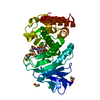
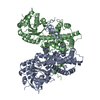
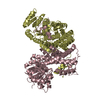
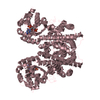
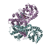
| Title | Dimeric 6-phosphogluconate dehydrogenase complexed with 6-phosphogluconate and 2'-monophosphoadenosine-5'-diphosphate | ||||||
 Components Components | 6-phosphogluconate dehydrogenase, decarboxylating | ||||||
 Keywords Keywords | OXIDOREDUCTASE / NADP / pentose phosphate pathway / 6-phosphogluconate dehydrogenase / 6-phosphogluconate / Gluconate utilization / Pentose shunt | ||||||
| Function / homology |  Function and homology information Function and homology informationphosphogluconate dehydrogenase (NADP+-dependent, decarboxylating) / D-gluconate metabolic process / phosphogluconate dehydrogenase (decarboxylating) activity / organic acid catabolic process / pentose-phosphate shunt, oxidative branch / pentose-phosphate shunt / guanosine tetraphosphate binding / NADP binding / protein homodimerization activity / identical protein binding / cytosol Similarity search - Function | ||||||
| Biological species |  | ||||||
| Method |  X-RAY DIFFRACTION / SYNCHROTRON / MOLECULAR REPLACEMENT / Resolution: 1.5 Å X-RAY DIFFRACTION / SYNCHROTRON / MOLECULAR REPLACEMENT / Resolution: 1.5 Å | ||||||
 Authors Authors | Chen, Y.-Y. / Ko, T.-P. / Lo, L.-P. / Lin, C.-H. / Wang, A.H.-J. | ||||||
 Citation Citation | Journal: J.Struct.Biol. / Year: 2010 Title: Conformational changes associated with cofactor/substrate binding of 6-phosphogluconate dehydrogenase from Escherichia coli and Klebsiella pneumoniae: Implications for enzyme mechanism Authors: Chen, Y.-Y. / Ko, T.-P. / Chen, W.-H. / Lo, L.-P. / Lin, C.-H. / Wang, A.H.-J. | ||||||
| History |
|
- Structure visualization
| Structure viewer | Molecule: MolmilJmol/JSmol |
|---|
- Downloads & links
-Download
| PDBx/mmCIF format | 3fwn.cif.gz | 234.6 KB | Display | PDBx/mmCIF format |
|---|---|---|---|---|
| PDB format | pdb3fwn.ent.gz | 182 KB | Display | PDB format |
| PDBx/mmJSON format | 3fwn.json.gz | Tree view | PDBx/mmJSON format | |
| Others |  Other downloads Other downloads |
-Validation report
| Summary document | 3fwn_validation.pdf.gz | 1.4 MB | Display | wwPDB validaton report |
|---|---|---|---|---|
| Full document | 3fwn_full_validation.pdf.gz | 1.5 MB | Display | |
| Data in XML | 3fwn_validation.xml.gz | 54.7 KB | Display | |
| Data in CIF | 3fwn_validation.cif.gz | 87.1 KB | Display | |
| Arichive directory | https://data.pdbj.org/pub/pdb/validation_reports/fw/3fwnftp://data.pdbj.org/pub/pdb/validation_reports/fw/3fwn | HTTPS FTP |
-Related structure data
| Related structure data |  2zyaC  2zydC 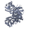 2zygC 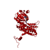 1pgoS C: citing same article ( S: Starting model for refinement |
|---|---|
| Similar structure data |


-Links
 PDBj
PDBj


- Assembly
| Deposited unit | 
| ||||||||
|---|---|---|---|---|---|---|---|---|---|
| 1 |
| ||||||||
| Unit cell |
|
-Components
| #1: Protein | Mass: 53101.809 Da / Num. of mol.: 2 / Mutation: N414I Source method: isolated from a genetically manipulated source Source: (gene. exp.) References: UniProt: P00350, phosphogluconate dehydrogenase (NADP+-dependent, decarboxylating) #2: Sugar |   Type: D-saccharide / Mass: 276.135 Da / Num. of mol.: 2 Type: D-saccharide / Mass: 276.135 Da / Num. of mol.: 2Source method: isolated from a genetically manipulated source Formula: C6H13O10P #3: Chemical | ChemComp-ATR / |   Mass: 507.181 Da / Num. of mol.: 1 / Source method: obtained synthetically / Formula: C10H16N5O13P3 Mass: 507.181 Da / Num. of mol.: 1 / Source method: obtained synthetically / Formula: C10H16N5O13P3#4: Water | ChemComp-HOH / |  Mass: 18.015 Da / Num. of mol.: 1681 / Source method: isolated from a natural source / Formula: H2O Mass: 18.015 Da / Num. of mol.: 1681 / Source method: isolated from a natural source / Formula: H2O |
|---|
-Experimental details
-Experiment
| Experiment | Method: X-RAY DIFFRACTION / Number of used crystals: 1 |
|---|
- Sample preparation
| Crystal | Density Matthews: 2.27 Å3/Da / Density % sol: 45.8 % |
|---|---|
| Crystal grow | Temperature: 291 K / Method: vapor diffusion, sitting drop / pH: 5.4 Details: 0.1M trisodium citrate dihrdrate, 0.5M ammonium acetate, 6-7 % PEG 3350, 17-18% PEG 4000, 3mM 6-phosphogluconate, 0.25mM NADPH, pH 5.4, VAPOR DIFFUSION, SITTING DROP, temperature 291K |
-Data collection
| Diffraction | Mean temperature: 100 K |
|---|---|
| Diffraction source | Source: SYNCHROTRON / Site: Photon Factory  / Beamline: BL-5A / Wavelength: 1 Å / Beamline: BL-5A / Wavelength: 1 Å |
| Detector | Type: ADSC QUANTUM 315 / Detector: CCD / Date: May 18, 2006 / Details: mirrors |
| Radiation | Monochromator: SAGITALLY FOCUSED Si(111) / Protocol: SINGLE WAVELENGTH / Monochromatic (M) / Laue (L): M / Scattering type: x-ray |
| Radiation wavelength | Wavelength: 1 Å / Relative weight: 1 |
| Reflection | Resolution: 1.5→50 Å / Num. all: 154920 / Num. obs: 150582 / % possible obs: 97.2 % / Redundancy: 6.8 % / Rmerge(I) obs: 0.068 / Net I/σ(I): 36.1 |
| Reflection shell | Resolution: 1.5→1.55 Å / Redundancy: 5.6 % / Rmerge(I) obs: 0.724 / Mean I/σ(I) obs: 2.7 / Num. unique all: 14395 / % possible all: 93.7 |
- Processing
| Software |
| |||||||||||||||||||||||||
|---|---|---|---|---|---|---|---|---|---|---|---|---|---|---|---|---|---|---|---|---|---|---|---|---|---|---|
| Refinement | Method to determine structure: MOLECULAR REPLACEMENT Starting model: 1pgo Resolution: 1.5→50 Å / Cross valid method: THROUGHOUT / σ(F): 0 / σ(I): 0 / Stereochemistry target values: Engh & Huber
| |||||||||||||||||||||||||
| Refine analyze |
| |||||||||||||||||||||||||
| Refinement step | Cycle: LAST / Resolution: 1.5→50 Å
| |||||||||||||||||||||||||
| Refine LS restraints |
| |||||||||||||||||||||||||
| LS refinement shell | Resolution: 1.5→1.55 Å / Rfactor Rfree error: 0.0309
|
