[English] 日本語
 Yorodumi
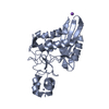
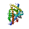
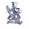
- PDB-3fvh: Polo-like kinase 1 Polo box domain in complex with Ac-LHSpTA-NH2 ... -
+ Open data
- Basic information
| Entry | Database: PDB / ID: 3fvh | ||||||
|---|---|---|---|---|---|---|---|
| Title | Polo-like kinase 1 Polo box domain in complex with Ac-LHSpTA-NH2 peptide | ||||||
 Components Components |
| ||||||
 Keywords Keywords | CELL CYCLE / PEPTIDE BINDING PROTEIN / Polo like kinase 1 / Polo box domain / phosphopeptide binding domain / ATP-binding / Cell division / Kinase / Mitosis / Nucleotide-binding / Nucleus / Phosphoprotein / Serine/threonine-protein kinase / Transferase | ||||||
| Function / homology |  Function and homology information Function and homology informationMitotic Telophase/Cytokinesis / regulation of protein localization to cell cortex / Mitotic Metaphase/Anaphase Transition / synaptonemal complex disassembly / Activation of NIMA Kinases NEK9, NEK6, NEK7 / polo kinase / mitotic nuclear membrane disassembly / Phosphorylation of Emi1 / protein localization to nuclear envelope / homologous chromosome segregation ...Mitotic Telophase/Cytokinesis / regulation of protein localization to cell cortex / Mitotic Metaphase/Anaphase Transition / synaptonemal complex disassembly / Activation of NIMA Kinases NEK9, NEK6, NEK7 / polo kinase / mitotic nuclear membrane disassembly / Phosphorylation of Emi1 / protein localization to nuclear envelope / homologous chromosome segregation / metaphase/anaphase transition of mitotic cell cycle / female meiosis chromosome segregation / nuclear membrane disassembly / synaptonemal complex / Phosphorylation of the APC/C / anaphase-promoting complex binding / Golgi inheritance / outer kinetochore / positive regulation of ubiquitin protein ligase activity / microtubule bundle formation / double-strand break repair via alternative nonhomologous end joining / mitotic chromosome condensation / Polo-like kinase mediated events / regulation of mitotic spindle assembly / Golgi Cisternae Pericentriolar Stack Reorganization / centrosome cycle / regulation of mitotic metaphase/anaphase transition / sister chromatid cohesion / positive regulation of ubiquitin-protein transferase activity / regulation of mitotic cell cycle phase transition / mitotic spindle assembly checkpoint signaling / mitotic spindle pole / spindle midzone / mitotic G2 DNA damage checkpoint signaling / regulation of anaphase-promoting complex-dependent catabolic process / mitotic cytokinesis / mitotic sister chromatid segregation / establishment of mitotic spindle orientation / positive regulation of proteolysis / negative regulation of double-strand break repair via homologous recombination / Regulation of MITF-M-dependent genes involved in cell cycle and proliferation / Cyclin A/B1/B2 associated events during G2/M transition / protein localization to chromatin / Amplification of signal from unattached kinetochores via a MAD2 inhibitory signal / Loss of Nlp from mitotic centrosomes / Loss of proteins required for interphase microtubule organization from the centrosome / Recruitment of mitotic centrosome proteins and complexes / centriole / Recruitment of NuMA to mitotic centrosomes / Mitotic Prometaphase / Anchoring of the basal body to the plasma membrane / EML4 and NUDC in mitotic spindle formation / regulation of mitotic cell cycle / AURKA Activation by TPX2 / Resolution of Sister Chromatid Cohesion / Condensation of Prophase Chromosomes / mitotic spindle organization / regulation of cytokinesis / establishment of protein localization / peptidyl-serine phosphorylation / RHO GTPases Activate Formins / APC/C:Cdh1 mediated degradation of Cdc20 and other APC/C:Cdh1 targeted proteins in late mitosis/early G1 / kinetochore / positive regulation of protein localization to nucleus / protein destabilization / G2/M transition of mitotic cell cycle / centriolar satellite / spindle / spindle pole / The role of GTSE1 in G2/M progression after G2 checkpoint / Separation of Sister Chromatids / Regulation of PLK1 Activity at G2/M Transition / mitotic cell cycle / double-strand break repair / positive regulation of proteasomal ubiquitin-dependent protein catabolic process / microtubule cytoskeleton / midbody / microtubule binding / protein phosphorylation / protein kinase activity / regulation of cell cycle / protein ubiquitination / protein serine kinase activity / protein serine/threonine kinase activity / centrosome / protein kinase binding / negative regulation of apoptotic process / chromatin / magnesium ion binding / negative regulation of transcription by RNA polymerase II / nucleoplasm / ATP binding / identical protein binding / nucleus / cytoplasm / cytosol Similarity search - Function | ||||||
| Biological species |  Homo sapiens (human) Homo sapiens (human) | ||||||
| Method |  X-RAY DIFFRACTION / X-RAY DIFFRACTION /  MOLECULAR REPLACEMENT / Resolution: 1.58 Å MOLECULAR REPLACEMENT / Resolution: 1.58 Å | ||||||
 Authors Authors | Lim, D.C. / Yaffe, M.B. | ||||||
 Citation Citation |  Journal: Nat.Struct.Mol.Biol. / Year: 2009 Journal: Nat.Struct.Mol.Biol. / Year: 2009Title: Structural and functional analyses of minimal phosphopeptides targeting the polo-box domain of polo-like kinase 1 Authors: Yun, S.M. / Moulaei, T. / Lim, D. / Bang, J.K. / Park, J.E. / Shenoy, S.R. / Liu, F. / Kang, Y.H. / Liao, C. / Soung, N.K. / Lee, S. / Yoon, D.Y. / Lim, Y. / Lee, D.H. / Otaka, A. / Appella, ...Authors: Yun, S.M. / Moulaei, T. / Lim, D. / Bang, J.K. / Park, J.E. / Shenoy, S.R. / Liu, F. / Kang, Y.H. / Liao, C. / Soung, N.K. / Lee, S. / Yoon, D.Y. / Lim, Y. / Lee, D.H. / Otaka, A. / Appella, E. / McMahon, J.B. / Nicklaus, M.C. / Burke, T.R. / Yaffe, M.B. / Wlodawer, A. / Lee, K.S. | ||||||
| History |
|
- Structure visualization
| Structure viewer | Molecule:  Molmil Molmil Jmol/JSmol Jmol/JSmol |
|---|
- Downloads & links
- Download
| PDBx/mmCIF format |  3fvh.cif.gz 3fvh.cif.gz | 108.2 KB | Display |  PDBx/mmCIF format PDBx/mmCIF format |
|---|---|---|---|---|
| PDB format |  pdb3fvh.ent.gz pdb3fvh.ent.gz | 83.4 KB | Display |  PDB format PDB format |
| PDBx/mmJSON format |  3fvh.json.gz 3fvh.json.gz | Tree view |  PDBx/mmJSON format PDBx/mmJSON format | |
| Others |  Other downloads Other downloads |
-Validation report
| Arichive directory |  https://data.pdbj.org/pub/pdb/validation_reports/fv/3fvh https://data.pdbj.org/pub/pdb/validation_reports/fv/3fvh ftp://data.pdbj.org/pub/pdb/validation_reports/fv/3fvh ftp://data.pdbj.org/pub/pdb/validation_reports/fv/3fvh | HTTPS FTP |
|---|
-Related structure data
| Related structure data |  3c5lC  3hihC  3hikC  1umwS S: Starting model for refinement C: citing same article ( |
|---|---|
| Similar structure data |
- Links
Links
- Assembly
| Deposited unit | 
| ||||||||
|---|---|---|---|---|---|---|---|---|---|
| 1 |
| ||||||||
| Unit cell |
|
- Components
| #1: Protein | Mass: 27285.158 Da / Num. of mol.: 1 Source method: isolated from a genetically manipulated source Source: (gene. exp.)  Homo sapiens (human) / Gene: PLK, PLK1 / Plasmid: pET28a / Production host: Homo sapiens (human) / Gene: PLK, PLK1 / Plasmid: pET28a / Production host:  |
|---|---|
| #2: Protein/peptide | Mass: 632.605 Da / Num. of mol.: 1 / Source method: obtained synthetically / Details: The peptide was chemically synthesized |
| #3: Water | ChemComp-HOH / |
| Has protein modification | Y |
-Experimental details
-Experiment
| Experiment | Method:  X-RAY DIFFRACTION / Number of used crystals: 1 X-RAY DIFFRACTION / Number of used crystals: 1 |
|---|
- Sample preparation
| Crystal | Density Matthews: 1.99 Å3/Da / Density % sol: 38.21 % |
|---|---|
| Crystal grow | Temperature: 293 K / Method: vapor diffusion, hanging drop / pH: 8.5 Details: PEG 2000 MME, Tris pH 8.5, trimethyl-amine oxide, VAPOR DIFFUSION, HANGING DROP, temperature 293K |
-Data collection
| Diffraction | Mean temperature: 100 K |
|---|---|
| Diffraction source | Source:  ROTATING ANODE / Type: RIGAKU MICROMAX-007 HF / Wavelength: 1.54 Å ROTATING ANODE / Type: RIGAKU MICROMAX-007 HF / Wavelength: 1.54 Å |
| Detector | Type: RIGAKU RAXIS IV++ / Detector: IMAGE PLATE / Date: Nov 8, 2008 / Details: Osmic |
| Radiation | Monochromator: Osmic mirrors / Protocol: SINGLE WAVELENGTH / Monochromatic (M) / Laue (L): M / Scattering type: x-ray |
| Radiation wavelength | Wavelength: 1.54 Å / Relative weight: 1 |
| Reflection | Resolution: 1.58→25 Å / Num. all: 30083 / Num. obs: 28949 / % possible obs: 96.4 % / Observed criterion σ(F): 2 / Observed criterion σ(I): 2 / Redundancy: 4.7 % / Biso Wilson estimate: 19.8 Å2 / Rsym value: 0.043 / Net I/σ(I): 17.4 |
| Reflection shell | Resolution: 1.58→1.64 Å / Redundancy: 4.5 % / Mean I/σ(I) obs: 8.6 / Num. unique all: 2834 / Rsym value: 0.172 / % possible all: 94.2 |
- Processing
| Software |
| |||||||||||||||||||||||||||||||||||||||||||||||||||||||||||||||||||||||||||||
|---|---|---|---|---|---|---|---|---|---|---|---|---|---|---|---|---|---|---|---|---|---|---|---|---|---|---|---|---|---|---|---|---|---|---|---|---|---|---|---|---|---|---|---|---|---|---|---|---|---|---|---|---|---|---|---|---|---|---|---|---|---|---|---|---|---|---|---|---|---|---|---|---|---|---|---|---|---|---|
| Refinement | Method to determine structure:  MOLECULAR REPLACEMENT MOLECULAR REPLACEMENTStarting model: PDB entry 1UMW Resolution: 1.58→24.171 Å / SU ML: 0.24 / Cross valid method: THROUGHOUT / σ(F): 1.32 / Stereochemistry target values: ML
| |||||||||||||||||||||||||||||||||||||||||||||||||||||||||||||||||||||||||||||
| Solvent computation | Shrinkage radii: 0.9 Å / VDW probe radii: 1.11 Å / Solvent model: FLAT BULK SOLVENT MODEL / Bsol: 48.962 Å2 / ksol: 0.395 e/Å3 | |||||||||||||||||||||||||||||||||||||||||||||||||||||||||||||||||||||||||||||
| Refinement step | Cycle: LAST / Resolution: 1.58→24.171 Å
| |||||||||||||||||||||||||||||||||||||||||||||||||||||||||||||||||||||||||||||
| LS refinement shell |
|
 Movie
Movie Controller


 PDBj
PDBj


