 Movie
Movie Controller


+ Open data

- Basic information
| Entry | Database: PDB / ID: 3fto | ||||||
|---|---|---|---|---|---|---|---|
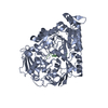
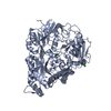
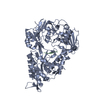
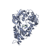
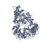
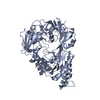
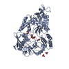
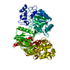
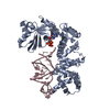
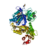
| Title | Crystal structure of OppA in a open conformation | ||||||
 Components Components | Oligopeptide-binding protein oppA | ||||||
 Keywords Keywords | PEPTIDE BINDING PROTEIN / oligo-peptide binding / voluminous binding cavity / venus fly-trap | ||||||
| Function / homology |  Function and homology information Function and homology informationpeptide transport / peptide transmembrane transporter activity / ATP-binding cassette (ABC) transporter complex / protein transport / periplasmic space Similarity search - Function | ||||||
| Biological species |  Lactococcus lactis subsp. cremoris (lactic acid bacteria) Lactococcus lactis subsp. cremoris (lactic acid bacteria) | ||||||
| Method |  X-RAY DIFFRACTION / SYNCHROTRON / SAD / Resolution: 2.38 Å X-RAY DIFFRACTION / SYNCHROTRON / SAD / Resolution: 2.38 Å | ||||||
 Authors Authors | Berntsson, R.P.-A. / Oktaviani, N.A. / Fusetti, F. / Thunnissen, A.-M.W.H. / Poolman, B. / Slotboom, D.-J. | ||||||
 Citation Citation | Journal: Protein Sci. / Year: 2009 Title: Selenomethionine incorporation in proteins expressed in Lactococcus lactis. Authors: Berntsson, R.P. / Alia Oktaviani, N. / Fusetti, F. / Thunnissen, A.M. / Poolman, B. / Slotboom, D.J. | ||||||
| History |
|
- Structure visualization
| Structure viewer | Molecule: MolmilJmol/JSmol |
|---|
- Downloads & links
-Download
| PDBx/mmCIF format | 3fto.cif.gz | 125.4 KB | Display | PDBx/mmCIF format |
|---|---|---|---|---|
| PDB format | pdb3fto.ent.gz | 95.2 KB | Display | PDB format |
| PDBx/mmJSON format | 3fto.json.gz | Tree view | PDBx/mmJSON format | |
| Others |  Other downloads Other downloads |
-Validation report
| Arichive directory | https://data.pdbj.org/pub/pdb/validation_reports/ft/3ftoftp://data.pdbj.org/pub/pdb/validation_reports/ft/3fto | HTTPS FTP |
|---|
-Related structure data
| Related structure data | |
|---|---|
| Similar structure data |


-Links
 PDBj
PDBj


- Assembly
| Deposited unit | 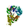
| ||||||||
|---|---|---|---|---|---|---|---|---|---|
| 1 |
| ||||||||
| Unit cell |
|
-Components
| #1: Protein | Mass: 65801.320 Da / Num. of mol.: 1 Source method: isolated from a genetically manipulated source Details: Construct lacking N-terminal signal sequence and cysteine used for lipid modification Source: (gene. exp.) Lactococcus lactis subsp. cremoris (lactic acid bacteria)Strain: MG1363 / Gene: llmg_0701, oppA / Plasmid: pAMP21 Production host: Lactococcus lactis subsp. cremoris (lactic acid bacteria)Strain (production host): MG1363 / References: UniProt: A2RJ53 |
|---|---|
| #2: Water | ChemComp-HOH /  Mass: 18.015 Da / Num. of mol.: 235 / Source method: isolated from a natural source / Formula: H2O Mass: 18.015 Da / Num. of mol.: 235 / Source method: isolated from a natural source / Formula: H2O |
| Has protein modification | Y |
-Experimental details
-Experiment
| Experiment | Method: X-RAY DIFFRACTION / Number of used crystals: 1 |
|---|
- Sample preparation
| Crystal | Density Matthews: 2.14 Å3/Da / Density % sol: 42.43 % / Mosaicity: 0.814 ° |
|---|---|
| Crystal grow | Temperature: 293 K / Method: vapor diffusion, hanging drop / pH: 7 Details: 0.2M NaCl, 0.1M Na-Hepes, 20% PEG 6000, pH 7.0, vapor diffusion, hanging drop, temperature 293K |
-Data collection
| Diffraction | Mean temperature: 100 K | |||||||||||||||||||||||||||||||||||||||||||||||||||||||||||||||||||||||||||||
|---|---|---|---|---|---|---|---|---|---|---|---|---|---|---|---|---|---|---|---|---|---|---|---|---|---|---|---|---|---|---|---|---|---|---|---|---|---|---|---|---|---|---|---|---|---|---|---|---|---|---|---|---|---|---|---|---|---|---|---|---|---|---|---|---|---|---|---|---|---|---|---|---|---|---|---|---|---|---|
| Diffraction source | Source: SYNCHROTRON / Site: ESRF  / Beamline: BM16 / Wavelength: 0.979 Å / Beamline: BM16 / Wavelength: 0.979 Å | |||||||||||||||||||||||||||||||||||||||||||||||||||||||||||||||||||||||||||||
| Detector | Type: ADSC Q210r / Detector: CCD / Date: Apr 20, 2008 | |||||||||||||||||||||||||||||||||||||||||||||||||||||||||||||||||||||||||||||
| Radiation | Protocol: SINGLE WAVELENGTH / Monochromatic (M) / Laue (L): M / Scattering type: x-ray | |||||||||||||||||||||||||||||||||||||||||||||||||||||||||||||||||||||||||||||
| Radiation wavelength | Wavelength: 0.979 Å / Relative weight: 1 | |||||||||||||||||||||||||||||||||||||||||||||||||||||||||||||||||||||||||||||
| Reflection | Redundancy: 6.2 % / Av σ(I) over netI: 44.89 / Number: 136650 / Rmerge(I) obs: 0.083 / Χ2: 4.59 / D res high: 2.38 Å / D res low: 50 Å / Num. obs: 22117 / % possible obs: 99.8 | |||||||||||||||||||||||||||||||||||||||||||||||||||||||||||||||||||||||||||||
| Diffraction reflection shell |
| |||||||||||||||||||||||||||||||||||||||||||||||||||||||||||||||||||||||||||||
| Reflection | Resolution: 2.38→50 Å / Num. obs: 22117 / % possible obs: 99.8 % / Redundancy: 6.2 % / Rmerge(I) obs: 0.083 / Χ2: 4.591 / Net I/σ(I): 44.886 | |||||||||||||||||||||||||||||||||||||||||||||||||||||||||||||||||||||||||||||
| Reflection shell |
|
-Phasing
| Phasing | Method: SAD | ||||||||||||||||||||||||||||||||||||||||||||||||||||||||||||||||||||||||||||||||||||||||||||||||||||||||||||||||
|---|---|---|---|---|---|---|---|---|---|---|---|---|---|---|---|---|---|---|---|---|---|---|---|---|---|---|---|---|---|---|---|---|---|---|---|---|---|---|---|---|---|---|---|---|---|---|---|---|---|---|---|---|---|---|---|---|---|---|---|---|---|---|---|---|---|---|---|---|---|---|---|---|---|---|---|---|---|---|---|---|---|---|---|---|---|---|---|---|---|---|---|---|---|---|---|---|---|---|---|---|---|---|---|---|---|---|---|---|---|---|---|---|---|
| Phasing MAD | D res high: 2.38 Å / D res low: 19.8 Å / FOM : 0.349 / FOM acentric: 0.359 / FOM centric: 0 / Reflection: 22071 / Reflection acentric: 21450 / Reflection centric: 621 | ||||||||||||||||||||||||||||||||||||||||||||||||||||||||||||||||||||||||||||||||||||||||||||||||||||||||||||||||
| Phasing MAD set | R cullis acentric: 1.68 / R cullis centric: 1 / Highest resolution: 2.38 Å / Lowest resolution: 19.8 Å / Loc acentric: 0.2 / Loc centric: 0.1 / Power acentric: 0 / Power centric: 0 / Reflection acentric: 21450 / Reflection centric: 621 | ||||||||||||||||||||||||||||||||||||||||||||||||||||||||||||||||||||||||||||||||||||||||||||||||||||||||||||||||
| Phasing MAD set shell | ID: 1 / R cullis centric: 1 / Power acentric: 0 / Power centric: 0
| ||||||||||||||||||||||||||||||||||||||||||||||||||||||||||||||||||||||||||||||||||||||||||||||||||||||||||||||||
| Phasing MAD set site |
| ||||||||||||||||||||||||||||||||||||||||||||||||||||||||||||||||||||||||||||||||||||||||||||||||||||||||||||||||
| Phasing MAD shell |
| ||||||||||||||||||||||||||||||||||||||||||||||||||||||||||||||||||||||||||||||||||||||||||||||||||||||||||||||||
| Phasing dm | Method: Solvent flattening and Histogram matching / Reflection: 22071 | ||||||||||||||||||||||||||||||||||||||||||||||||||||||||||||||||||||||||||||||||||||||||||||||||||||||||||||||||
| Phasing dm shell |
|
- Processing
| Software |
| |||||||||||||||||||||||||||||||||||||||||||||||||||||||||||||||||||||||||||||||||||||
|---|---|---|---|---|---|---|---|---|---|---|---|---|---|---|---|---|---|---|---|---|---|---|---|---|---|---|---|---|---|---|---|---|---|---|---|---|---|---|---|---|---|---|---|---|---|---|---|---|---|---|---|---|---|---|---|---|---|---|---|---|---|---|---|---|---|---|---|---|---|---|---|---|---|---|---|---|---|---|---|---|---|---|---|---|---|---|
| Refinement | Method to determine structure: SAD / Resolution: 2.38→21.23 Å / Cor.coef. Fo:Fc: 0.924 / Cor.coef. Fo:Fc free: 0.861 / WRfactor Rfree: 0.281 / WRfactor Rwork: 0.215 / Occupancy max: 1 / Occupancy min: 0.9 / FOM work R set: 0.806 / SU B: 8.967 / SU ML: 0.213 / SU R Cruickshank DPI: 0.713 / SU Rfree: 0.315 / Cross valid method: THROUGHOUT / σ(F): 0 / ESU R: 0.713 / ESU R Free: 0.315 / Stereochemistry target values: MAXIMUM LIKELIHOOD Details: HYDROGENS HAVE BEEN ADDED IN THE RIDING POSITIONS U VALUES: REFINED INDIVIDUALLY
| |||||||||||||||||||||||||||||||||||||||||||||||||||||||||||||||||||||||||||||||||||||
| Solvent computation | Ion probe radii: 0.8 Å / Shrinkage radii: 0.8 Å / VDW probe radii: 1.2 Å / Solvent model: BABINET MODEL WITH MASK | |||||||||||||||||||||||||||||||||||||||||||||||||||||||||||||||||||||||||||||||||||||
| Displacement parameters | Biso max: 68.39 Å2 / Biso mean: 35.666 Å2 / Biso min: 9.25 Å2
| |||||||||||||||||||||||||||||||||||||||||||||||||||||||||||||||||||||||||||||||||||||
| Refinement step | Cycle: LAST / Resolution: 2.38→21.23 Å
| |||||||||||||||||||||||||||||||||||||||||||||||||||||||||||||||||||||||||||||||||||||
| Refine LS restraints |
| |||||||||||||||||||||||||||||||||||||||||||||||||||||||||||||||||||||||||||||||||||||
| LS refinement shell | Resolution: 2.38→2.44 Å / Total num. of bins used: 20
|
