 Movie
Movie Controller

[English] 日本語
 Yorodumi
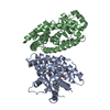
- PDB-3dri: Crystal structure of Lactococcal OppA co-crystallized with an oct... -
+ Open data

- Basic information
| Entry | Database: PDB / ID: 3dri | ||||||
|---|---|---|---|---|---|---|---|
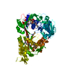
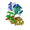
| Title | Crystal structure of Lactococcal OppA co-crystallized with an octamer peptide in an open conformation | ||||||
 Components Components |
| ||||||
 Keywords Keywords | PEPTIDE BINDING PROTEIN / oligo-peptide binding / voluminous binding cavity / venus fly-trap | ||||||
| Function / homology |  Function and homology information Function and homology informationpeptide transport / peptide transmembrane transporter activity / ATP-binding cassette (ABC) transporter complex / protein transport / periplasmic space Similarity search - Function | ||||||
| Biological species |  Lactococcus lactis (lactic acid bacteria) Lactococcus lactis (lactic acid bacteria) | ||||||
| Method |  X-RAY DIFFRACTION / SYNCHROTRON / MOLECULAR REPLACEMENT / molecular replacement / Resolution: 1.8 Å X-RAY DIFFRACTION / SYNCHROTRON / MOLECULAR REPLACEMENT / molecular replacement / Resolution: 1.8 Å | ||||||
 Authors Authors | Berntsson, R.P.-A. / Doeven, M.K. / Duurkens, R.H. / Sengupta, D. / Marrink, S.-J. / Thunnissen, A.-M. / Poolman, B. / Slotboom, D.-J. | ||||||
 Citation Citation | Journal: Embo J. / Year: 2009 Title: The structural basis for peptide selection by the transport receptor OppA Authors: Berntsson, R.P.-A. / Doeven, M.K. / Fusetti, F. / Duurkens, R.H. / Sengupta, D. / Marrink, S.-J. / Thunnissen, A.-M. / Poolman, B. / Slotboom, D.-J. | ||||||
| History |
|
- Structure visualization
| Structure viewer | Molecule: MolmilJmol/JSmol |
|---|
- Downloads & links
-Download
| PDBx/mmCIF format | 3dri.cif.gz | 140.6 KB | Display | PDBx/mmCIF format |
|---|---|---|---|---|
| PDB format | pdb3dri.ent.gz | 106.1 KB | Display | PDB format |
| PDBx/mmJSON format | 3dri.json.gz | Tree view | PDBx/mmJSON format | |
| Others |  Other downloads Other downloads |
-Validation report
| Arichive directory | https://data.pdbj.org/pub/pdb/validation_reports/dr/3driftp://data.pdbj.org/pub/pdb/validation_reports/dr/3dri | HTTPS FTP |
|---|
-Related structure data
| Related structure data | 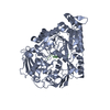 3drfSC 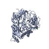 3drgC 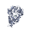 3drhC 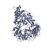 3drjC  3drkC S: Starting model for refinement C: citing same article ( |
|---|---|
| Similar structure data |


-Links
 PDBj
PDBj


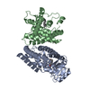
- Assembly
| Deposited unit | 
| ||||||||
|---|---|---|---|---|---|---|---|---|---|
| 1 |
| ||||||||
| Unit cell |
|
-Components
| #1: Protein | Mass: 65144.773 Da / Num. of mol.: 1 Source method: isolated from a genetically manipulated source Details: Construct lacking N-terminal signal sequence and cysteine used for lipid modification Source: (gene. exp.) Lactococcus lactis (lactic acid bacteria)Strain: subsp. cremoris MG1363 / Gene: oppA, llmg_0701 / Plasmid: pAMP21 / Production host: Lactococcus lactis (lactic acid bacteria) / Strain (production host): MG1363 / References: UniProt: A2RJ53 |
|---|---|
| #2: Protein/peptide | Mass: 476.482 Da / Num. of mol.: 1 / Source method: obtained synthetically / Details: chemically synthesis |
| #3: Water | ChemComp-HOH /  Mass: 18.015 Da / Num. of mol.: 774 / Source method: isolated from a natural source / Formula: H2O Mass: 18.015 Da / Num. of mol.: 774 / Source method: isolated from a natural source / Formula: H2O |
-Experimental details
-Experiment
| Experiment | Method: X-RAY DIFFRACTION / Number of used crystals: 1 |
|---|
- Sample preparation
| Crystal | Density Matthews: 2.17 Å3/Da / Density % sol: 43.39 % / Mosaicity: 0.57 ° |
|---|---|
| Crystal grow | Temperature: 293 K / Method: vapor diffusion, hanging drop / pH: 7 Details: 0.2M NaCl, 0.1M Na-Hepes, 20% PEG 6000, pH 7.0, vapor diffusion, hanging drop, temperature 293K |
-Data collection
| Diffraction | Mean temperature: 100 K | ||||||||||||||||||||||||||||||||||||||||||||||||||||||||||||||||||||||||||||||||||||||||
|---|---|---|---|---|---|---|---|---|---|---|---|---|---|---|---|---|---|---|---|---|---|---|---|---|---|---|---|---|---|---|---|---|---|---|---|---|---|---|---|---|---|---|---|---|---|---|---|---|---|---|---|---|---|---|---|---|---|---|---|---|---|---|---|---|---|---|---|---|---|---|---|---|---|---|---|---|---|---|---|---|---|---|---|---|---|---|---|---|---|
| Diffraction source | Source: SYNCHROTRON / Site: ESRF  / Beamline: ID14-2 / Wavelength: 0.931 Å / Beamline: ID14-2 / Wavelength: 0.931 Å | ||||||||||||||||||||||||||||||||||||||||||||||||||||||||||||||||||||||||||||||||||||||||
| Detector | Type: ADSC Q210 / Detector: CCD / Date: Sep 22, 2007 | ||||||||||||||||||||||||||||||||||||||||||||||||||||||||||||||||||||||||||||||||||||||||
| Radiation | Protocol: SINGLE WAVELENGTH / Monochromatic (M) / Laue (L): M / Scattering type: x-ray | ||||||||||||||||||||||||||||||||||||||||||||||||||||||||||||||||||||||||||||||||||||||||
| Radiation wavelength | Wavelength: 0.931 Å / Relative weight: 1 | ||||||||||||||||||||||||||||||||||||||||||||||||||||||||||||||||||||||||||||||||||||||||
| Reflection | Redundancy: 3.7 % / Av σ(I) over netI: 2.3 / Number: 192649 / Rmerge(I) obs: 0.108 / Rsym value: 0.108 / D res high: 1.8 Å / D res low: 61.663 Å / Num. obs: 51727 / % possible obs: 100 | ||||||||||||||||||||||||||||||||||||||||||||||||||||||||||||||||||||||||||||||||||||||||
| Diffraction reflection shell |
| ||||||||||||||||||||||||||||||||||||||||||||||||||||||||||||||||||||||||||||||||||||||||
| Reflection | Resolution: 1.8→61.663 Å / Num. obs: 51727 / % possible obs: 100 % / Redundancy: 3.7 % / Rmerge(I) obs: 0.108 / Rsym value: 0.108 / Net I/σ(I): 2.3 | ||||||||||||||||||||||||||||||||||||||||||||||||||||||||||||||||||||||||||||||||||||||||
| Reflection shell | Diffraction-ID: 1
|
-Phasing
| Phasing | Method: molecular replacement | |||||||||
|---|---|---|---|---|---|---|---|---|---|---|
| Phasing MR | Model details: Phaser MODE: MR_AUTO
|
- Processing
| Software |
| ||||||||||||||||||||||||||||||||||||||||||||||||||||||||||||||||||||||||||||||||||||||||||||||||||||
|---|---|---|---|---|---|---|---|---|---|---|---|---|---|---|---|---|---|---|---|---|---|---|---|---|---|---|---|---|---|---|---|---|---|---|---|---|---|---|---|---|---|---|---|---|---|---|---|---|---|---|---|---|---|---|---|---|---|---|---|---|---|---|---|---|---|---|---|---|---|---|---|---|---|---|---|---|---|---|---|---|---|---|---|---|---|---|---|---|---|---|---|---|---|---|---|---|---|---|---|---|---|
| Refinement | Method to determine structure: MOLECULAR REPLACEMENT Starting model: 3DRF Resolution: 1.8→42.37 Å / Cor.coef. Fo:Fc: 0.958 / Cor.coef. Fo:Fc free: 0.938 / SU B: 4.805 / SU ML: 0.082 / TLS residual ADP flag: LIKELY RESIDUAL / Cross valid method: THROUGHOUT / σ(F): 0 / ESU R: 0.126 / ESU R Free: 0.121 / Stereochemistry target values: MAXIMUM LIKELIHOOD
| ||||||||||||||||||||||||||||||||||||||||||||||||||||||||||||||||||||||||||||||||||||||||||||||||||||
| Solvent computation | Ion probe radii: 0.8 Å / Shrinkage radii: 0.8 Å / VDW probe radii: 1.2 Å / Solvent model: BABINET MODEL WITH MASK | ||||||||||||||||||||||||||||||||||||||||||||||||||||||||||||||||||||||||||||||||||||||||||||||||||||
| Displacement parameters | Biso mean: 14.571 Å2
| ||||||||||||||||||||||||||||||||||||||||||||||||||||||||||||||||||||||||||||||||||||||||||||||||||||
| Refinement step | Cycle: LAST / Resolution: 1.8→42.37 Å
| ||||||||||||||||||||||||||||||||||||||||||||||||||||||||||||||||||||||||||||||||||||||||||||||||||||
| Refine LS restraints |
| ||||||||||||||||||||||||||||||||||||||||||||||||||||||||||||||||||||||||||||||||||||||||||||||||||||
| LS refinement shell | Resolution: 1.8→1.847 Å / Total num. of bins used: 20
| ||||||||||||||||||||||||||||||||||||||||||||||||||||||||||||||||||||||||||||||||||||||||||||||||||||
| Refinement TLS params. | Method: refined / Refine-ID: X-RAY DIFFRACTION
| ||||||||||||||||||||||||||||||||||||||||||||||||||||||||||||||||||||||||||||||||||||||||||||||||||||
| Refinement TLS group |
|
