 Movie
Movie Controller

[English] 日本語
 Yorodumi
- PDB-3fsh: Crystal structure of the ubiquitin conjugating enzyme Ube2g2 boun... -
+ Open data

- Basic information
| Entry | Database: PDB / ID: 3fsh | ||||||
|---|---|---|---|---|---|---|---|
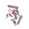
| Title | Crystal structure of the ubiquitin conjugating enzyme Ube2g2 bound to the G2BR domain of ubiquitin ligase gp78 | ||||||
 Components Components |
| ||||||
 Keywords Keywords | LIGASE / PROTEIN-PEPTIDE COMPLEX / Ubl conjugation pathway / Alternative splicing / Endoplasmic reticulum / Membrane / Metal-binding / Phosphoprotein / Polymorphism / Receptor / Transmembrane / Zinc / Zinc-finger | ||||||
| Function / homology |  Function and homology information Function and homology informationregulation of SREBP signaling pathway / negative regulation of retrograde protein transport, ER to cytosol / RING-type E3 ubiquitin transferase (cysteine targeting) / endoplasmic reticulum mannose trimming / Synthesis of active ubiquitin: roles of E1 and E2 enzymes / endoplasmic reticulum quality control compartment / BAT3 complex binding / Derlin-1 retrotranslocation complex / Antigen processing: Ubiquitination & Proteasome degradation / protein K27-linked ubiquitination ...regulation of SREBP signaling pathway / negative regulation of retrograde protein transport, ER to cytosol / RING-type E3 ubiquitin transferase (cysteine targeting) / endoplasmic reticulum mannose trimming / Synthesis of active ubiquitin: roles of E1 and E2 enzymes / endoplasmic reticulum quality control compartment / BAT3 complex binding / Derlin-1 retrotranslocation complex / Antigen processing: Ubiquitination & Proteasome degradation / protein K27-linked ubiquitination / E2 ubiquitin-conjugating enzyme / non-canonical NF-kappaB signal transduction / ubiquitin-ubiquitin ligase activity / ubiquitin-specific protease binding / ubiquitin conjugating enzyme activity / ubiquitin ligase complex / protein autoubiquitination / cellular response to interferon-beta / endoplasmic reticulum unfolded protein response / ERAD pathway / protein K48-linked ubiquitination / lipid droplet / ER Quality Control Compartment (ERQC) / protein catabolic process / ubiquitin binding / N-glycan trimming in the ER and Calnexin/Calreticulin cycle / negative regulation of canonical Wnt signaling pathway / Wnt signaling pathway / protein polyubiquitination / ubiquitin-protein transferase activity / ubiquitin protein ligase activity / growth cone / protein-folding chaperone binding / signaling receptor activity / ubiquitin-dependent protein catabolic process / learning or memory / protein-macromolecule adaptor activity / neuronal cell body / dendrite / endoplasmic reticulum membrane / perinuclear region of cytoplasm / Golgi apparatus / endoplasmic reticulum / signal transduction / protein-containing complex / zinc ion binding / ATP binding / membrane / identical protein binding / cytosol Similarity search - Function | ||||||
| Biological species |  | ||||||
| Method |  X-RAY DIFFRACTION / SYNCHROTRON / MOLECULAR REPLACEMENT / Resolution: 2.76 Å X-RAY DIFFRACTION / SYNCHROTRON / MOLECULAR REPLACEMENT / Resolution: 2.76 Å | ||||||
 Authors Authors | Tu, D. / Brunger, A.T. | ||||||
 Citation Citation | Journal: Proc.Natl.Acad.Sci.USA / Year: 2009 Title: Mechanistic insights into active site-associated polyubiquitination by the ubiquitin-conjugating enzyme Ube2g2. Authors: Li, W. / Tu, D. / Li, L. / Wollert, T. / Ghirlando, R. / Brunger, A.T. / Ye, Y. | ||||||
| History |
|
- Structure visualization
| Structure viewer | Molecule: MolmilJmol/JSmol |
|---|
- Downloads & links
-Download
| PDBx/mmCIF format | 3fsh.cif.gz | 82.4 KB | Display | PDBx/mmCIF format |
|---|---|---|---|---|
| PDB format | pdb3fsh.ent.gz | 62.2 KB | Display | PDB format |
| PDBx/mmJSON format | 3fsh.json.gz | Tree view | PDBx/mmJSON format | |
| Others |  Other downloads Other downloads |
-Validation report
| Arichive directory | https://data.pdbj.org/pub/pdb/validation_reports/fs/3fshftp://data.pdbj.org/pub/pdb/validation_reports/fs/3fsh | HTTPS FTP |
|---|
-Related structure data
| Related structure data | 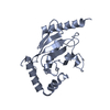 2cyxS S: Starting model for refinement |
|---|---|
| Similar structure data |


-Links
 PDBj
PDBj


- Assembly
| Deposited unit | 
| ||||||||
|---|---|---|---|---|---|---|---|---|---|
| 1 | 
| ||||||||
| 2 | 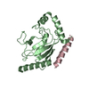
| ||||||||
| Unit cell |
|
-Components
| #1: Protein | Mass: 18864.537 Da / Num. of mol.: 2 Source method: isolated from a genetically manipulated source Source: (gene. exp.)  #2: Protein/peptide | | Mass: 3452.988 Da / Num. of mol.: 1 / Fragment: G2BR / Source method: obtained synthetically Details: THE PEPTIDE WAS CHEMICALLY SYNTHESIZED. THIS SEQUENCE OCCURS NATURALLY IN HUMANS. References: UniProt: Q9UKV5, Ligases; Forming carbon-nitrogen bonds; Acid-amino-acid ligases (peptide synthases) #3: Water | ChemComp-HOH / |  Mass: 18.015 Da / Num. of mol.: 40 / Source method: isolated from a natural source / Formula: H2O Mass: 18.015 Da / Num. of mol.: 40 / Source method: isolated from a natural source / Formula: H2O |
|---|
-Experimental details
-Experiment
| Experiment | Method: X-RAY DIFFRACTION / Number of used crystals: 1 |
|---|
- Sample preparation
| Crystal | Density Matthews: 4.84 Å3/Da / Density % sol: 74.6 % |
|---|---|
| Crystal grow | pH: 7.7 Details: 2.8M SODIUM FORMATE, 0.1M TRIS PH 7.7, 5MM DTT, VAPOR DIFFUSION, HANGING DROP, TEMPERATURE 293K |
-Data collection
| Diffraction | Mean temperature: 100 K |
|---|---|
| Diffraction source | Source: SYNCHROTRON / Site: SSRL  / Beamline: BL11-1 / Wavelength: 0.97945 / Beamline: BL11-1 / Wavelength: 0.97945 |
| Detector | Type: MARMOSAIC 325 mm CCD / Detector: CCD / Date: Feb 27, 2007 |
| Radiation | Monochromator: FLAT MIRROR(VERTICAL FOCUSING), SINGLE CRYSTAL SI(111) BENT MONOCHROMATOR (HORIZONTAL FOCUSING) Protocol: SINGLE WAVELENGTH / Monochromatic (M) / Laue (L): M / Scattering type: x-ray |
| Radiation wavelength | Wavelength: 0.97945 Å / Relative weight: 1 |
| Reflection | Resolution: 2.76→50 Å / Num. obs: 21654 / % possible obs: 99.5 % / Observed criterion σ(I): -3 / Redundancy: 8.4 % / Biso Wilson estimate: 53.1 Å2 / Rsym value: 0.105 / Net I/σ(I): 19.4 |
| Reflection shell | Resolution: 2.76→2.86 Å / Redundancy: 5.8 % / Mean I/σ(I) obs: 2.1 / Rsym value: 0.65 / % possible all: 95.5 |
- Processing
| Software |
| ||||||||||||||||||||||||||||||||||||||||||||||||||||||||||||
|---|---|---|---|---|---|---|---|---|---|---|---|---|---|---|---|---|---|---|---|---|---|---|---|---|---|---|---|---|---|---|---|---|---|---|---|---|---|---|---|---|---|---|---|---|---|---|---|---|---|---|---|---|---|---|---|---|---|---|---|---|---|
| Refinement | Method to determine structure: MOLECULAR REPLACEMENT Starting model: PDB ENTRY 2CYX Resolution: 2.76→50 Å / Isotropic thermal model: ISOTROPIC / Cross valid method: THROUGHOUT / σ(F): 0 / Stereochemistry target values: ENGH & HUBER
| ||||||||||||||||||||||||||||||||||||||||||||||||||||||||||||
| Displacement parameters | Biso mean: 61.28 Å2 | ||||||||||||||||||||||||||||||||||||||||||||||||||||||||||||
| Refine analyze |
| ||||||||||||||||||||||||||||||||||||||||||||||||||||||||||||
| Refinement step | Cycle: LAST / Resolution: 2.76→50 Å
| ||||||||||||||||||||||||||||||||||||||||||||||||||||||||||||
| Refine LS restraints |
| ||||||||||||||||||||||||||||||||||||||||||||||||||||||||||||
| LS refinement shell | Resolution: 2.76→2.86 Å / Total num. of bins used: 10
|
