[English] 日本語
Yorodumi
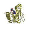
- PDB-3fmb: Crystal structure of dimeric protein of unknown function and ferr... -

+
Open data


ID or keywords:

Loading...

-
Basic information

Entry
Database: PDB / ID: 3fmb
TitleCrystal structure of dimeric protein of unknown function and ferredoxin-like fold (YP_212648.1) from Bacteroides fragilis NCTC 9343 at 1.85 A resolution
ComponentsDimeric protein of unknown function and ferredoxin-like fold
KeywordsSTRUCTURAL GENOMICS / UNKNOWN FUNCTION / YP_212648.1 / Stress responsive A/B Barrel Domain / dimeric protein of unknown function and ferredoxin-like fold / Joint Center for Structural Genomics / JCSG / Protein Structure Initiative / PSI-2
Function / homology
Function and homology information


Stress responsive alpha-beta barrel / Stress responsive A/B Barrel Domain / Stress-response A/B barrel domain profile. / Stress responsive A/B Barrel Domain / Alpha-Beta Plaits - #100 / Dimeric alpha-beta barrel / Alpha-Beta Plaits / 2-Layer Sandwich / Alpha Beta
Similarity search - Domain/homology
Stress-response A/B barrel domain-containing protein
Similarity search - Component
Biological speciesBacteroides fragilis (bacteria)
MethodX-RAY DIFFRACTION / SYNCHROTRON / MAD / Resolution: 1.85 Å
AuthorsJoint Center for Structural Genomics (JCSG)
CitationJournal: To be published
Title: Crystal structure of dimeric protein of unknown function and ferredoxin-like fold (YP_212648.1) from Bacteroides fragilis NCTC 9343 at 1.85 A resolution
Authors: Joint Center for Structural Genomics (JCSG)
History
DepositionDec 19, 2008Deposition site: RCSB / Processing site: RCSB
Revision 1.0Jan 27, 2009Provider: repository / Type: Initial release
Revision 1.1Jul 13, 2011Group: Advisory / Source and taxonomy / Version format compliance
Revision 1.2Nov 1, 2017Group: Refinement description / Category: software / Item: _software.classification / _software.name
Revision 1.3Jul 24, 2019Group: Data collection / Derived calculations / Refinement description
Category: software / struct_conn
Item: _software.classification / _software.contact_author ..._software.classification / _software.contact_author / _software.contact_author_email / _software.language / _software.location / _software.name / _software.type / _software.version / _struct_conn.pdbx_leaving_atom_flag
Revision 1.4Feb 1, 2023Group: Database references / Derived calculations / Category: database_2 / struct_ref_seq_dif / struct_site
Item: _database_2.pdbx_DOI / _database_2.pdbx_database_accession ..._database_2.pdbx_DOI / _database_2.pdbx_database_accession / _struct_ref_seq_dif.details / _struct_site.pdbx_auth_asym_id / _struct_site.pdbx_auth_comp_id / _struct_site.pdbx_auth_seq_id

-
Structure visualization

Structure viewerMolecule:
MolmilJmol/JSmol

Downloads & links

-
Assembly

Deposited unit
A: Dimeric protein of unknown function and ferredoxin-like fold
B: Dimeric protein of unknown function and ferredoxin-like fold
hetero molecules


Theoretical massNumber of molelcules
Total (without water)28,0959
Polymers27,5922
Non-polymers5027
Water5,296294
1


  • Idetical with deposited unit
  • defined by author&software
TypeNameSymmetry operationNumber
identity operation1_555x,y,z1
Buried area2230 Å2
ΔGint-15.6 kcal/mol
Surface area10340 Å2
MethodPISA
Unit cell
Length a, b, c (Å)72.310, 72.310, 233.020
Angle α, β, γ (deg.)90.000, 90.000, 120.000
Int Tables number178
Space group name H-MP6122
Noncrystallographic symmetry (NCS)NCS domain:
IDEns-IDDetails
11A
21B

NCS domain segments:
Dom-IDComponent-IDEns-IDRefine codeAuth asym-IDAuth seq-ID
1116A-3 - 99
2116B-3 - 99

-
Components

#1: Protein Dimeric protein of unknown function and ferredoxin-like fold


Mass: 13796.136 Da / Num. of mol.: 2
Source method: isolated from a genetically manipulated source
Source: (gene. exp.) Bacteroides fragilis (bacteria) / Strain: NCTC 9343 / Gene: BF3034, YP_212648.1 / Plasmid: SpeedET / Production host: Escherichia coli (E. coli) / Strain (production host): HK100 / References: UniProt: Q5LAZ3
#2: Chemical ChemComp-SO4 / SULFATE ION


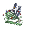
Mass: 96.063 Da / Num. of mol.: 2 / Source method: obtained synthetically / Formula: SO4
#3: Chemical
ChemComp-EDO / 1,2-ETHANEDIOL / ETHYLENE GLYCOL


Mass: 62.068 Da / Num. of mol.: 5 / Source method: obtained synthetically / Formula: C2H6O2
#4: Water ChemComp-HOH / water


Mass: 18.015 Da / Num. of mol.: 294 / Source method: isolated from a natural source / Formula: H2O
Sequence detailsTHE CONSTRUCT WAS EXPRESSED WITH A PURIFICATION TAG MGSDKIHHHHHHENLYFQG.

-
Experimental details

-
Experiment

ExperimentMethod: X-RAY DIFFRACTION / Number of used crystals: 1

-
Sample preparation

CrystalDensity Matthews: 3.19 Å3/Da / Density % sol: 61.4 %
Crystal growTemperature: 277 K / Method: vapor diffusion, sitting drop / pH: 6
Details: NANODROP, 0.2M (NH4)2SO4, 20.0% PEG 3350, No Buffer pH 6.0, VAPOR DIFFUSION, SITTING DROP, temperature 277K

-
Data collection

DiffractionMean temperature: 100 K
Diffraction sourceSource: SYNCHROTRON / Site: SSRL / Beamline: BL11-1 / Wavelength: 0.91837, 0.97867
DetectorType: MARMOSAIC 325 mm CCD / Detector: CCD / Date: Nov 12, 2008 / Details: Flat mirror (vertical focusing)
RadiationMonochromator: Single crystal Si(111) bent (horizontal focusing)
Protocol: MAD / Monochromatic (M) / Laue (L): M / Scattering type: x-ray
Radiation wavelength
IDWavelength (Å)Relative weight
10.918371
20.978671
ReflectionResolution: 1.85→29.399 Å / Num. obs: 31933 / % possible obs: 99.7 % / Observed criterion σ(I): -3 / Biso Wilson estimate: 29.156 Å2 / Rmerge(I) obs: 0.09
Reflection shell
Resolution (Å)Rmerge(I) obsMean I/σ(I) obsNum. measured obsNum. unique obsDiffraction-ID% possible all
1.85-1.920.0121.7390826033198.6
1.92-1.990.0122.5364165322199.8
1.99-2.080.0123.64048858201100
2.08-2.190.0125.24188458961100
2.19-2.330.0126.8432425948199.9
2.33-2.510.0129.6434955833199.9
2.51-2.760.01213.7442815783199.9
2.76-3.160.01220453625859199.9
3.16-3.980.01231.9450945849199.8
3.98-29.3990.01239.3453905884199.5

-
Phasing

PhasingMethod: MAD

-
Processing

Software
NameVersionClassificationNB
REFMAC5.2.0019refinement
PHENIXrefinement
MolProbity3beta29model building
XSCALEdata scaling
PDB_EXTRACT3.006data extraction
MAR345CCDdata collection
XDSdata reduction
SHELXCDphasing
autoSHARPphasing
RefinementMethod to determine structure: MAD / Resolution: 1.85→29.399 Å / Cor.coef. Fo:Fc: 0.965 / Cor.coef. Fo:Fc free: 0.951 / Occupancy max: 1 / Occupancy min: 0.37 / SU B: 4.322 / SU ML: 0.068 / TLS residual ADP flag: LIKELY RESIDUAL / Cross valid method: THROUGHOUT / σ(F): 0 / ESU R: 0.098 / ESU R Free: 0.1
Stereochemistry target values: MAXIMUM LIKELIHOOD WITH PHASES
Details: 1. HYDROGENS HAVE BEEN ADDED IN THE RIDING POSITIONS. 2. ATOM RECORDS CONTAIN RESIDUAL B FACTORS ONLY. 3. A MET-INHIBITION PROTOCOL WAS USED FOR SELENOMETHIONINE INCORPORATION DURING PROTEIN ...Details: 1. HYDROGENS HAVE BEEN ADDED IN THE RIDING POSITIONS. 2. ATOM RECORDS CONTAIN RESIDUAL B FACTORS ONLY. 3. A MET-INHIBITION PROTOCOL WAS USED FOR SELENOMETHIONINE INCORPORATION DURING PROTEIN EXPRESSION. THE OCCUPANCY OF THE SE ATOMS IN THE MSE RESIDUES WAS REDUCED TO 0.75 FOR THE REDUCED SCATTERING POWER DUE TO PARTIAL S-MET INCORPORATION. 4. SULFATE (SO4) IONS FROM CRYSTALLIZATION CONDITION AND ETHYLENE GLYCOL (EDO) FROM CRYO SOLUTION WERE MODELED.
RfactorNum. reflection% reflectionSelection details
Rfree0.205 1612 5.1 %RANDOM
Rwork0.17 ---
obs0.172 31830 99.72 %-
Solvent computationIon probe radii: 0.8 Å / Shrinkage radii: 0.8 Å / VDW probe radii: 1.2 Å / Solvent model: MASK
Displacement parametersBiso max: 76.73 Å2 / Biso mean: 32.402 Å2 / Biso min: 18.59 Å2
Baniso -1Baniso -2Baniso -3
1-0.58 Å20.29 Å20 Å2
2--0.58 Å20 Å2
3----0.87 Å2
Refinement stepCycle: LAST / Resolution: 1.85→29.399 Å
ProteinNucleic acidLigandSolventTotal
Num. atoms1646 0 30 294 1970
Refine LS restraints
Refine-IDTypeDev idealDev ideal targetNumber
X-RAY DIFFRACTIONr_bond_refined_d0.0170.0221780
X-RAY DIFFRACTIONr_bond_other_d0.0080.021212
X-RAY DIFFRACTIONr_angle_refined_deg1.4681.9722407
X-RAY DIFFRACTIONr_angle_other_deg0.97132975
X-RAY DIFFRACTIONr_dihedral_angle_1_deg5.5455220
X-RAY DIFFRACTIONr_dihedral_angle_2_deg31.69725.12282
X-RAY DIFFRACTIONr_dihedral_angle_3_deg13.02815322
X-RAY DIFFRACTIONr_dihedral_angle_4_deg14.104157
X-RAY DIFFRACTIONr_chiral_restr0.0930.2264
X-RAY DIFFRACTIONr_gen_planes_refined0.0070.021975
X-RAY DIFFRACTIONr_gen_planes_other0.0020.02346
X-RAY DIFFRACTIONr_nbd_refined0.2120.2335
X-RAY DIFFRACTIONr_nbd_other0.1990.21251
X-RAY DIFFRACTIONr_nbtor_refined0.1760.2845
X-RAY DIFFRACTIONr_nbtor_other0.0850.2878
X-RAY DIFFRACTIONr_xyhbond_nbd_refined0.1670.2220
X-RAY DIFFRACTIONr_symmetry_vdw_refined0.1480.210
X-RAY DIFFRACTIONr_symmetry_vdw_other0.1850.221
X-RAY DIFFRACTIONr_symmetry_hbond_refined0.1860.211
X-RAY DIFFRACTIONr_mcbond_it2.0831336
X-RAY DIFFRACTIONr_mcbond_other0.5083426
X-RAY DIFFRACTIONr_mcangle_it2.4451756
X-RAY DIFFRACTIONr_scbond_it4.7318796
X-RAY DIFFRACTIONr_scangle_it6.46511651
Refine LS restraints NCS

Auth asym-ID: A / Ens-ID: 1 / Number: 1349 / Refine-ID: X-RAY DIFFRACTION

Dom-IDTypeRms dev position (Å)Weight position
1LOOSE POSITIONAL0.355
2LOOSE THERMAL1.7610
LS refinement shellResolution: 1.85→1.897 Å / Total num. of bins used: 20
RfactorNum. reflection% reflection
Rfree0.358 110 -
Rwork0.261 2135 -
all-2245 -
obs--97.91 %
Refinement TLS params.

Method: refined / Refine-ID: X-RAY DIFFRACTION

IDL112)L122)L132)L222)L232)L332)S11 (Å °)S12 (Å °)S13 (Å °)S21 (Å °)S22 (Å °)S23 (Å °)S31 (Å °)S32 (Å °)S33 (Å °)T112)T122)T132)T222)T232)T332)Origin x (Å)Origin y (Å)Origin z (Å)
12.29730.68490.33930.98930.25162.83660.0283-0.1668-0.06980.1071-0.05850.03160.34040.03720.0302-0.0857-0.02190.0425-0.0544-0.0009-0.0607-7.037821.641810.0772
22.4509-0.2267-0.02890.7210.26332.6405-0.00550.31390.0295-0.047-0.02640.00740.0470.05810.032-0.1463-0.02210.02350.0068-0.0063-0.0651-9.005626.7741-7.4316
Refinement TLS group
IDRefine-IDRefine TLS-IDAuth asym-IDAuth seq-ID
1X-RAY DIFFRACTION1A1 - 99
2X-RAY DIFFRACTION2B2 - 99

+
About Yorodumi

-
News

-
Feb 9, 2022. New format data for meta-information of EMDB entries

New format data for meta-information of EMDB entries

  • Version 3 of the EMDB header file is now the official format.
  • The previous official version 1.9 will be removed from the archive.

Related info.:EMDB header

External links:wwPDB to switch to version 3 of the EMDB data model

-
Aug 12, 2020. Covid-19 info

Covid-19 info

URL: https://pdbjlvh1.pdbj.org/emnavi/covid19.php

New page: Covid-19 featured information page in EM Navigator.

Related info.:Covid-19 info / Mar 5, 2020. Novel coronavirus structure data

+
Mar 5, 2020. Novel coronavirus structure data

Novel coronavirus structure data

Related info.:Yorodumi Speices / Aug 12, 2020. Covid-19 info

External links:COVID-19 featured content - PDBj / Molecule of the Month (242):Coronavirus Proteases

+
Jan 31, 2019. EMDB accession codes are about to change! (news from PDBe EMDB page)

EMDB accession codes are about to change! (news from PDBe EMDB page)

  • The allocation of 4 digits for EMDB accession codes will soon come to an end. Whilst these codes will remain in use, new EMDB accession codes will include an additional digit and will expand incrementally as the available range of codes is exhausted. The current 4-digit format prefixed with “EMD-” (i.e. EMD-XXXX) will advance to a 5-digit format (i.e. EMD-XXXXX), and so on. It is currently estimated that the 4-digit codes will be depleted around Spring 2019, at which point the 5-digit format will come into force.
  • The EM Navigator/Yorodumi systems omit the EMD- prefix.

Related info.:Q: What is EMD? / ID/Accession-code notation in Yorodumi/EM Navigator

External links:EMDB Accession Codes are Changing Soon! / Contact to PDBj

+
Jul 12, 2017. Major update of PDB

Major update of PDB

  • wwPDB released updated PDB data conforming to the new PDBx/mmCIF dictionary.
  • This is a major update changing the version number from 4 to 5, and with Remediation, in which all the entries are updated.
  • In this update, many items about electron microscopy experimental information are reorganized (e.g. em_software).
  • Now, EM Navigator and Yorodumi are based on the updated data.

External links:wwPDB Remediation / Enriched Model Files Conforming to OneDep Data Standards Now Available in the PDB FTP Archive

-
Yorodumi

Thousand views of thousand structures

  • Yorodumi is a browser for structure data from EMDB, PDB, SASBDB, etc.
  • This page is also the successor to EM Navigator detail page, and also detail information page/front-end page for Omokage search.
  • The word "yorodu" (or yorozu) is an old Japanese word meaning "ten thousand". "mi" (miru) is to see.

Related info.:EMDB / PDB / SASBDB / Comparison of 3 databanks / Yorodumi Search / Aug 31, 2016. New EM Navigator & Yorodumi / Yorodumi Papers / Jmol/JSmol / Function and homology information / Changes in new EM Navigator and Yorodumi

Read more