 Movie
Movie Controller

[English] 日本語
 Yorodumi
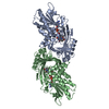
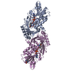
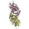
- PDB-3fb6: KcsA Potassium channel in the partially open state with 16 A open... -
+ Open data

- Basic information
| Entry | Database: PDB / ID: 3fb6 | ||||||
|---|---|---|---|---|---|---|---|
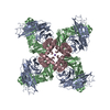
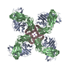
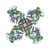
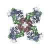
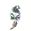
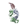
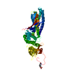
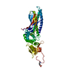
| Title | KcsA Potassium channel in the partially open state with 16 A opening at T112 | ||||||
 Components Components |
| ||||||
 Keywords Keywords | membrane protein/metal transport / kcsa / open / inactivation / potassium channel / Cell membrane / Ion transport / Ionic channel / Membrane / Transmembrane / Transport / Voltage-gated channel / membrane protein-metal transport COMPLEX | ||||||
| Function / homology |  Function and homology information Function and homology informationalpha-beta T cell receptor complex / IgG immunoglobulin complex / action potential / voltage-gated potassium channel activity / voltage-gated potassium channel complex / B cell differentiation / adaptive immune response / extracellular region / identical protein binding / plasma membrane Similarity search - Function | ||||||
| Biological species |  Streptomyces lividans (bacteria) Streptomyces lividans (bacteria) | ||||||
| Method |  X-RAY DIFFRACTION / SYNCHROTRON / MOLECULAR REPLACEMENT / Resolution: 3 Å X-RAY DIFFRACTION / SYNCHROTRON / MOLECULAR REPLACEMENT / Resolution: 3 Å | ||||||
 Authors Authors | Cuello, L.G. / Jogini, V. / Cortes, D.M. / Perozo, E. | ||||||
 Citation Citation | Journal: TO BE PUBLISHED Title: KcsA Potassium channel in the partially open state with 16 A opening at T112 Authors: Cuello, L.G. / Jogini, V. / Cortes, D.M. / Perozo, E. | ||||||
| History |
|
- Structure visualization
| Structure viewer | Molecule: MolmilJmol/JSmol |
|---|
- Downloads & links
-Download
| PDBx/mmCIF format | 3fb6.cif.gz | 110.7 KB | Display | PDBx/mmCIF format |
|---|---|---|---|---|
| PDB format | pdb3fb6.ent.gz | 85 KB | Display | PDB format |
| PDBx/mmJSON format | 3fb6.json.gz | Tree view | PDBx/mmJSON format | |
| Others |  Other downloads Other downloads |
-Validation report
| Arichive directory | https://data.pdbj.org/pub/pdb/validation_reports/fb/3fb6ftp://data.pdbj.org/pub/pdb/validation_reports/fb/3fb6 | HTTPS FTP |
|---|
-Related structure data
| Related structure data |  1k4cS S: Starting model for refinement |
|---|---|
| Similar structure data |


-Links
 PDBj
PDBj


- Assembly
| Deposited unit | 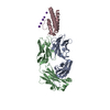
| ||||||||||||||||||
|---|---|---|---|---|---|---|---|---|---|---|---|---|---|---|---|---|---|---|---|
| 1 |
| ||||||||||||||||||
| 2 | 
| ||||||||||||||||||
| Unit cell |
| ||||||||||||||||||
| Components on special symmetry positions |
|
-Components
| #1: Antibody | Mass: 23411.242 Da / Num. of mol.: 1 / Source method: isolated from a natural source / Source: (natural) | ||
|---|---|---|---|
| #2: Antibody | Mass: 23435.738 Da / Num. of mol.: 1 / Source method: isolated from a natural source / Source: (natural) | ||
| #3: Protein | Mass: 10927.571 Da / Num. of mol.: 1 / Fragment: UNP residues 21-124 / Mutation: H25Q,L90C,R117Q,E120Q,R121Q,R122Q,H124Q Source method: isolated from a genetically manipulated source Source: (gene. exp.) Streptomyces lividans (bacteria) / Gene: kcsA, skc1 / Production host: | ||
| #4: Chemical | ChemComp-K /   Mass: 39.098 Da / Num. of mol.: 5 / Source method: obtained synthetically / Formula: K Mass: 39.098 Da / Num. of mol.: 5 / Source method: obtained synthetically / Formula: KHas protein modification | Y | |
-Experimental details
-Experiment
| Experiment | Method: X-RAY DIFFRACTION / Number of used crystals: 1 |
|---|
- Sample preparation
| Crystal | Density Matthews: 3.89 Å3/Da / Density % sol: 68.35 % |
|---|---|
| Crystal grow | Temperature: 298 K / Method: vapor diffusion, sitting drop Details: 20-25% PEG400, 50mM magnesium acetate, 50mM sodium acetate pH 5.0-6.0, pH 5-6, VAPOR DIFFUSION, SITTING DROP, temperature 298K PH range: 5-6 |
-Data collection
| Diffraction source | Source: SYNCHROTRON / Site: APS  / Beamline: 24-ID-C / Wavelength: 1 Å / Beamline: 24-ID-C / Wavelength: 1 Å |
|---|---|
| Detector | Type: ADSC QUANTUM 315 / Detector: CCD / Date: Oct 13, 2006 |
| Radiation | Protocol: SINGLE WAVELENGTH / Monochromatic (M) / Laue (L): M / Scattering type: x-ray |
| Radiation wavelength | Wavelength: 1 Å / Relative weight: 1 |
| Reflection | Resolution: 3→40 Å / Num. all: 17949 / Num. obs: 17088 / % possible obs: 95.2 % / Observed criterion σ(F): 2 / Observed criterion σ(I): 2 |
- Processing
| Software |
| ||||||||||||||||||
|---|---|---|---|---|---|---|---|---|---|---|---|---|---|---|---|---|---|---|---|
| Refinement | Method to determine structure: MOLECULAR REPLACEMENT Starting model: PDB entry 1K4C Resolution: 3→40 Å / σ(F): 2
| ||||||||||||||||||
| Refinement step | Cycle: LAST / Resolution: 3→40 Å
| ||||||||||||||||||
| Refine LS restraints |
|
