 Movie
Movie Controller


+ Open data

- Basic information
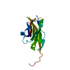
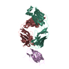
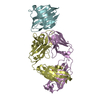
| Entry | Database: PDB / ID: 1pkq | ||||||
|---|---|---|---|---|---|---|---|
| Title | Myelin Oligodendrocyte Glycoprotein-(8-18C5) Fab-complex | ||||||
 Components Components |
| ||||||
 Keywords Keywords | IMMUNE SYSTEM / autoantibody / multiple sclerosis / EAE | ||||||
| Function / homology |  Function and homology information Function and homology informationresponse to folic acid / response to lipid / regulation of cytokine production / T cell receptor signaling pathway / cell adhesion / external side of plasma membrane / signaling receptor binding / cell surface Similarity search - Function | ||||||
| Biological species |  | ||||||
| Method |  X-RAY DIFFRACTION / SYNCHROTRON / MOLECULAR REPLACEMENT / Resolution: 3 Å X-RAY DIFFRACTION / SYNCHROTRON / MOLECULAR REPLACEMENT / Resolution: 3 Å | ||||||
 Authors Authors | Breithaupt, C. / Schubart, A. / Zander, H. / Skerra, A. / Huber, R. / Linington, C. / Jacob, U. | ||||||
 Citation Citation | Journal: Proc.Natl.Acad.Sci.USA / Year: 2003 Title: Structural insights into the antigenicity of myelin oligodendrocyte glycoprotein Authors: Breithaupt, C. / Schubart, A. / Zander, H. / Skerra, A. / Huber, R. / Linington, C. / Jacob, U. | ||||||
| History |
|
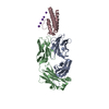
- Structure visualization
| Structure viewer | Molecule: MolmilJmol/JSmol |
|---|
- Downloads & links
-Download
| PDBx/mmCIF format | 1pkq.cif.gz | 224.2 KB | Display | PDBx/mmCIF format |
|---|---|---|---|---|
| PDB format | pdb1pkq.ent.gz | 177.3 KB | Display | PDB format |
| PDBx/mmJSON format | 1pkq.json.gz | Tree view | PDBx/mmJSON format | |
| Others |  Other downloads Other downloads |
-Validation report
| Arichive directory | https://data.pdbj.org/pub/pdb/validation_reports/pk/1pkqftp://data.pdbj.org/pub/pdb/validation_reports/pk/1pkq | HTTPS FTP |
|---|
-Related structure data


-Links
 PDBj
PDBj


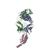
- Assembly
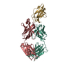
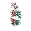
| Deposited unit | 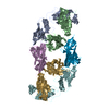
| ||||||||
|---|---|---|---|---|---|---|---|---|---|
| 1 | 
| ||||||||
| 2 | 
| ||||||||
| Unit cell |
|
-Components
| #1: Antibody | Mass: 26154.316 Da / Num. of mol.: 2 Source method: isolated from a genetically manipulated source Source: (gene. exp.)  #2: Antibody | Mass: 26683.082 Da / Num. of mol.: 2 Source method: isolated from a genetically manipulated source Source: (gene. exp.) #3: Protein | Mass: 15928.754 Da / Num. of mol.: 2 / Fragment: N-terminal Ig domain Source method: isolated from a genetically manipulated source Source: (gene. exp.) #4: Water | ChemComp-HOH / |  Mass: 18.015 Da / Num. of mol.: 85 / Source method: isolated from a natural source / Formula: H2O Mass: 18.015 Da / Num. of mol.: 85 / Source method: isolated from a natural source / Formula: H2OHas protein modification | Y | |
|---|
-Experimental details
-Experiment
| Experiment | Method: X-RAY DIFFRACTION / Number of used crystals: 1 |
|---|
- Sample preparation
| Crystal | Density Matthews: 2.67 Å3/Da / Density % sol: 53.55 % | ||||||||||||||||||||||||||||||||||||||||||
|---|---|---|---|---|---|---|---|---|---|---|---|---|---|---|---|---|---|---|---|---|---|---|---|---|---|---|---|---|---|---|---|---|---|---|---|---|---|---|---|---|---|---|---|
| Crystal grow | Temperature: 291 K / Method: vapor diffusion, sitting drop / pH: 8.5 Details: Tris/HCl, MOPS, NaCl, PEG8000, pH 8.5, VAPOR DIFFUSION, SITTING DROP, temperature 291K | ||||||||||||||||||||||||||||||||||||||||||
| Crystal grow | *PLUS Temperature: 18 ℃ / pH: 7.5 / Method: vapor diffusion, sitting drop | ||||||||||||||||||||||||||||||||||||||||||
| Components of the solutions | *PLUS
|
-Data collection
| Diffraction | Mean temperature: 100 K |
|---|---|
| Diffraction source | Source: SYNCHROTRON / Site: MPG/DESY, HAMBURG  / Beamline: BW6 / Wavelength: 1.05 Å / Beamline: BW6 / Wavelength: 1.05 Å |
| Detector | Type: MARRESEARCH / Detector: CCD / Date: Nov 10, 2002 |
| Radiation | Monochromator: Si(111) double crystal, non dispersive / Protocol: SINGLE WAVELENGTH / Monochromatic (M) / Laue (L): M / Scattering type: x-ray |
| Radiation wavelength | Wavelength: 1.05 Å / Relative weight: 1 |
| Reflection | Resolution: 3→18 Å / Num. obs: 22442 / % possible obs: 86.7 % / Redundancy: 2.3 % / Biso Wilson estimate: 69.1 Å2 / Rmerge(I) obs: 0.094 / Net I/σ(I): 5.3 |
| Reflection shell | Resolution: 3→3.09 Å / Redundancy: 1.9 % / Rmerge(I) obs: 0.351 / Mean I/σ(I) obs: 2 / Num. unique all: 1590 / % possible all: 73 |
| Reflection | *PLUS Num. obs: 22490 |
| Reflection shell | *PLUS Highest resolution: 3 Å / Lowest resolution: 3.1 Å / % possible obs: 73 % |
- Processing
| Software |
| |||||||||||||||||||||||||
|---|---|---|---|---|---|---|---|---|---|---|---|---|---|---|---|---|---|---|---|---|---|---|---|---|---|---|
| Refinement | Method to determine structure: MOLECULAR REPLACEMENT / Resolution: 3→18 Å / Cross valid method: THROUGHOUT / σ(F): 0 / Stereochemistry target values: Engh & Huber
| |||||||||||||||||||||||||
| Displacement parameters | Biso mean: 33.6 Å2 | |||||||||||||||||||||||||
| Refinement step | Cycle: LAST / Resolution: 3→18 Å
| |||||||||||||||||||||||||
| Refine LS restraints |
| |||||||||||||||||||||||||
| LS refinement shell | Resolution: 3→3.05 Å /
| |||||||||||||||||||||||||
| Refinement | *PLUS Rfactor Rwork: 0.25 | |||||||||||||||||||||||||
| Solvent computation | *PLUS | |||||||||||||||||||||||||
| Displacement parameters | *PLUS |
