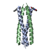
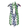
 Movie
Movie Controller

[English] 日本語
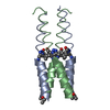
 Yorodumi
- PDB-3f87: An alpha/beta-Peptide Helix Bundle with a Pure beta-Amino Acid Co... -
+ Open data

- Basic information
| Entry | Database: PDB / ID: 3f87 | |||||||||
|---|---|---|---|---|---|---|---|---|---|---|
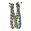
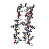
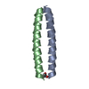
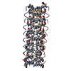
| Title | An alpha/beta-Peptide Helix Bundle with a Pure beta-Amino Acid Core and a Distinctive Quarternary Structure: GCN4pLI derivative with beta residues at a and d heptad positions - higher symmetry crystal | |||||||||
 Components Components | GCN4pLI-betaAD | |||||||||
 Keywords Keywords | UNKNOWN FUNCTION / alpha/beta-peptide / helix bundle / foldamer / GCN4 derivative | |||||||||
| Function / homology | IODIDE ION Function and homology information Function and homology information | |||||||||
| Method |  X-RAY DIFFRACTION / MOLECULAR REPLACEMENT / Resolution: 2.4 Å X-RAY DIFFRACTION / MOLECULAR REPLACEMENT / Resolution: 2.4 Å | |||||||||
 Authors Authors | Giuliano, M.W. / Horne, W.S. / Gellman, S.H. | |||||||||
 Citation Citation | Journal: J.Am.Chem.Soc. / Year: 2009 Title: An alpha/beta-peptide helix bundle with a pure beta3-amino acid core and a distinctive quaternary structure. Authors: Giuliano, M.W. / Horne, W.S. / Gellman, S.H. | |||||||||
| History |
|
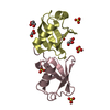
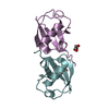
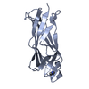
- Structure visualization
| Structure viewer | Molecule: MolmilJmol/JSmol |
|---|
- Downloads & links
-Download
| PDBx/mmCIF format | 3f87.cif.gz | 73.6 KB | Display | PDBx/mmCIF format |
|---|---|---|---|---|
| PDB format | pdb3f87.ent.gz | 64.1 KB | Display | PDB format |
| PDBx/mmJSON format | 3f87.json.gz | Tree view | PDBx/mmJSON format | |
| Others |  Other downloads Other downloads |
-Validation report
| Arichive directory | https://data.pdbj.org/pub/pdb/validation_reports/f8/3f87ftp://data.pdbj.org/pub/pdb/validation_reports/f8/3f87 | HTTPS FTP |
|---|
-Related structure data


-Links
 PDBj
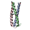
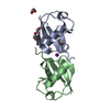
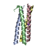
PDBj- Assembly
| Deposited unit | 
| ||||||||
|---|---|---|---|---|---|---|---|---|---|
| 1 | 
| ||||||||
| 2 | 
| ||||||||
| Unit cell |
| ||||||||
| Details | The asymmetric unit contains half of two separate, NCS-related tetrameric helix bundles: The second half of the first tetramer, which contains chains A and B, is generated by: (-y, -x, -z+1/2) + (010) / The asymmetric unit contains half of two separate, NCS-related tetrameric helix bundles: The second half of the second tetramer, which contains chains C and D, is generated by: (y, x, -z) + (100) |
-Components
| #1: Protein/peptide | Mass: 4212.099 Da / Num. of mol.: 4 / Source method: obtained synthetically / Details: Synthetic Peptide / References: PDB-3F86#2: Chemical | ChemComp-IOD /   Mass: 126.904 Da / Num. of mol.: 9 / Source method: obtained synthetically / Formula: I Mass: 126.904 Da / Num. of mol.: 9 / Source method: obtained synthetically / Formula: I#3: Water | ChemComp-HOH / |  Mass: 18.015 Da / Num. of mol.: 79 / Source method: isolated from a natural source / Formula: H2O Mass: 18.015 Da / Num. of mol.: 79 / Source method: isolated from a natural source / Formula: H2O |
|---|
-Experimental details
-Experiment
| Experiment | Method: X-RAY DIFFRACTION / Number of used crystals: 1 |
|---|
- Sample preparation
| Crystal | Density Matthews: 3.68 Å3/Da / Density % sol: 66.54 % |
|---|---|
| Crystal grow | Temperature: 298 K / Method: vapor diffusion, hanging drop / pH: 7.5 Details: 0.12 M NaI, 0.08 M NaCl, 0.1 M HEPES-Na pH 7.5, 20% (v/v) 2-methyl-2,4-pentanediol (MPD), VAPOR DIFFUSION, HANGING DROP, temperature 298K |
-Data collection
| Diffraction | Mean temperature: 100 K |
|---|---|
| Diffraction source | Source: ROTATING ANODE / Type: BRUKER AXS MICROSTAR / Wavelength: 1.5418 Å |
| Detector | Type: BRUKER SMART 6000 / Detector: CCD / Date: Jun 21, 2007 / Details: confocal mirrors |
| Radiation | Monochromator: Gobel mirrors / Protocol: SINGLE WAVELENGTH / Monochromatic (M) / Laue (L): M / Scattering type: x-ray |
| Radiation wavelength | Wavelength: 1.5418 Å / Relative weight: 1 |
| Reflection | Resolution: 2.4→50.4 Å / Num. all: 10451 / Num. obs: 10316 / % possible obs: 98.7 % / Redundancy: 12.61 % / Rmerge(I) obs: 0.148 / Rsym value: 0.061 |
| Reflection shell | Resolution: 2.4→2.49 Å / Redundancy: 9.76 % / Rmerge(I) obs: 0.521 / Mean I/σ(I) obs: 4.41 / Num. unique all: 1096 / Rsym value: 0.228 / % possible all: 99.5 |
- Processing
| Software |
| |||||||||||||||||||||||||||||||||||||||||||||||||||||||||||||||||||||||||||||||||||||
|---|---|---|---|---|---|---|---|---|---|---|---|---|---|---|---|---|---|---|---|---|---|---|---|---|---|---|---|---|---|---|---|---|---|---|---|---|---|---|---|---|---|---|---|---|---|---|---|---|---|---|---|---|---|---|---|---|---|---|---|---|---|---|---|---|---|---|---|---|---|---|---|---|---|---|---|---|---|---|---|---|---|---|---|---|---|---|
| Refinement | Method to determine structure: MOLECULAR REPLACEMENT / Resolution: 2.4→50.4 Å / Cor.coef. Fo:Fc: 0.925 / Cor.coef. Fo:Fc free: 0.884 / SU B: 7.794 / SU ML: 0.182 / Cross valid method: THROUGHOUT / ESU R: 0.298 / ESU R Free: 0.263 / Stereochemistry target values: MAXIMUM LIKELIHOOD / Details: HYDROGENS HAVE BEEN ADDED IN THE RIDING POSITIONS
| |||||||||||||||||||||||||||||||||||||||||||||||||||||||||||||||||||||||||||||||||||||
| Solvent computation | Ion probe radii: 0.8 Å / Shrinkage radii: 0.8 Å / VDW probe radii: 1.4 Å / Solvent model: MASK | |||||||||||||||||||||||||||||||||||||||||||||||||||||||||||||||||||||||||||||||||||||
| Displacement parameters | Biso mean: 17.868 Å2
| |||||||||||||||||||||||||||||||||||||||||||||||||||||||||||||||||||||||||||||||||||||
| Refinement step | Cycle: LAST / Resolution: 2.4→50.4 Å
| |||||||||||||||||||||||||||||||||||||||||||||||||||||||||||||||||||||||||||||||||||||
| Refine LS restraints |
| |||||||||||||||||||||||||||||||||||||||||||||||||||||||||||||||||||||||||||||||||||||
| LS refinement shell | Resolution: 2.396→2.458 Å / Total num. of bins used: 20
|
