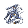
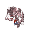
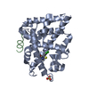
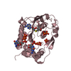
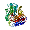
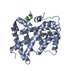
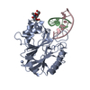
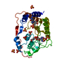
[English] 日本語
Yorodumi
- PDB-3f7d: SF-1 LBD bound by phosphatidylcholine -

+
Open data


ID or keywords:

Loading...

-
Basic information

Entry
Database: PDB / ID: 3f7d
TitleSF-1 LBD bound by phosphatidylcholine
Components
  • Peroxisome proliferator-activated receptor gamma coactivator 1-alphaPeroxisome proliferator-activated receptor gamma
  • nuclear receptor SF-1
KeywordsTRANSCRIPTION / nuclear receptor / coactivator peptide / ligand / phospholipid / phosphatidylcholine / transcriptional regulation / DNA-binding / Metal-binding / Nucleus / Receptor / Transcription regulation / Zinc-finger / Activator / RNA-binding
Function / homology
Function and homology information


flavone metabolic process / : / : / positive regulation of cellular metabolic process / response to metformin / response to norepinephrine / : / euchromatin => GO:0000791 / cellular response to resveratrol / cellular response to nitrite ...flavone metabolic process / : / : / positive regulation of cellular metabolic process / response to metformin / response to norepinephrine / : / euchromatin => GO:0000791 / cellular response to resveratrol / cellular response to nitrite / response to gonadotropin-releasing hormone / positive regulation of progesterone biosynthetic process / Sertoli cell differentiation / response to electrical stimulus involved in regulation of muscle adaptation / reproductive process / : / cellular response to ionomycin / SUMOylation of intracellular receptors / negative regulation of female gonad development / Nuclear Receptor transcription pathway / positive regulation of mitochondrial DNA metabolic process / positive regulation of muscle tissue development / skeletal muscle atrophy / sex determination / response to methionine / positive regulation of male gonad development / positive regulation of podocyte apoptotic process / negative regulation of smooth muscle cell migration / luteinization / adaptive thermogenesis / calcineurin-mediated signaling / : / positive regulation of cellular respiration / response to xenobiotic stimulus => GO:0009410 / response to epinephrine / positive regulation of fatty acid oxidation / tissue development / chromatin => GO:0000785 / cellular response to fructose stimulus / cellular response to follicle-stimulating hormone stimulus / response to L-leucine / cellular response to potassium ion / autophagy of mitochondrion / Leydig cell differentiation / galactose metabolic process / : / male sex determination / apical dendrite / maintenance of protein location in nucleus / cellular response to interleukin-6 / hormone metabolic process / cellular response to thyroid hormone stimulus / negative regulation of glycolytic process / peroxisome proliferator activated receptor binding / response to muscle activity / adrenal gland development / female gonad development / positive regulation of ATP biosynthetic process / response to starvation / cellular response to caffeine / fatty acid oxidation / regulation of NMDA receptor activity / response to dietary excess / transcription factor binding / alpha-tubulin binding / androgen metabolic process / negative regulation of mitochondrial fission / adipose tissue development / energy homeostasis / brown fat cell differentiation / forebrain development / negative regulation of signaling receptor activity / cellular response to transforming growth factor beta stimulus / respiratory electron transport chain / response to cold / hormone-mediated signaling pathway / cerebellum development / mitochondrion organization / cytosolic ribosome / response to reactive oxygen species / nuclear receptor coactivator activity / negative regulation of protein phosphorylation / response to activity / cellular response to estradiol stimulus / response to ischemia / gluconeogenesis / promoter-specific chromatin binding / transcription coregulator binding / nuclear estrogen receptor binding / nuclear receptor binding / cellular response to glucose stimulus / transcription coregulator activity / negative regulation of smooth muscle cell proliferation / RNA polymerase II transcription regulatory region sequence-specific DNA binding / positive regulation of smooth muscle cell proliferation / phospholipid binding / circadian regulation of gene expression / regulation of circadian rhythm / PML body / chromatin DNA binding
Similarity search - Function
Peroxisome proliferator-activated receptor gamma coactivator 1-alpha / PGC-1alpha, RNA recognition motif / PGC-1 / Nuclear hormone receptor family 5 / Retinoid X Receptor / Retinoid X Receptor / RNA recognition motif / RNA recognition motif / Eukaryotic RNA Recognition Motif (RRM) profile. / RNA recognition motif domain ...Peroxisome proliferator-activated receptor gamma coactivator 1-alpha / PGC-1alpha, RNA recognition motif / PGC-1 / Nuclear hormone receptor family 5 / Retinoid X Receptor / Retinoid X Receptor / RNA recognition motif / RNA recognition motif / Eukaryotic RNA Recognition Motif (RRM) profile. / RNA recognition motif domain / RNA-binding domain superfamily / Nuclear hormone receptor / Nuclear hormones receptors DNA-binding region signature. / Zinc finger, nuclear hormone receptor-type / Zinc finger, C4 type (two domains) / Nuclear hormone receptors DNA-binding domain profile. / c4 zinc finger in nuclear hormone receptors / Nuclear hormone receptor, ligand-binding domain / Nuclear hormone receptor-like domain superfamily / Ligand-binding domain of nuclear hormone receptor / Nuclear receptor (NR) ligand-binding (LBD) domain profile. / Ligand binding domain of hormone receptors / Zinc finger, NHR/GATA-type / Nucleotide-binding alpha-beta plait domain superfamily / Orthogonal Bundle / Mainly Alpha
Similarity search - Domain/homology
Chem-P42 / Peroxisome proliferator-activated receptor gamma coactivator 1-alpha / Steroidogenic factor 1 / Steroidogenic factor 1
Similarity search - Component
Biological speciesMus musculus (house mouse)
MethodX-RAY DIFFRACTION / SYNCHROTRON / MOLECULAR REPLACEMENT / Resolution: 2.2 Å
AuthorsSablin, E.P. / Fletterick, R.J.
CitationJournal: Mol.Endocrinol. / Year: 2009
Title: Structure of SF-1 bound by different phospholipids: evidence for regulatory ligands.
Authors: Sablin, E.P. / Blind, R.D. / Krylova, I.N. / Ingraham, J.G. / Cai, F. / Williams, J.D. / Fletterick, R.J. / Ingraham, H.A.
History
DepositionNov 7, 2008Deposition site: RCSB / Processing site: RCSB
Revision 1.0Dec 9, 2008Provider: repository / Type: Initial release
Revision 1.1Jul 13, 2011Group: Version format compliance
Revision 1.2Apr 2, 2014Group: Source and taxonomy
Revision 1.3Jul 24, 2019Group: Data collection / Derived calculations / Refinement description
Category: software / struct_conn
Item: _software.classification / _software.name ..._software.classification / _software.name / _software.version / _struct_conn.pdbx_leaving_atom_flag
Revision 1.4Oct 20, 2021Group: Database references / Derived calculations / Category: database_2 / struct_ref_seq_dif / struct_site
Item: _database_2.pdbx_DOI / _database_2.pdbx_database_accession ..._database_2.pdbx_DOI / _database_2.pdbx_database_accession / _struct_ref_seq_dif.details / _struct_site.pdbx_auth_asym_id / _struct_site.pdbx_auth_comp_id / _struct_site.pdbx_auth_seq_id
Revision 1.5Sep 6, 2023Group: Data collection / Refinement description
Category: chem_comp_atom / chem_comp_bond / pdbx_initial_refinement_model

-
Structure visualization

Structure viewerMolecule:
MolmilJmol/JSmol

Downloads & links

-
Assembly

Deposited unit
A: nuclear receptor SF-1
B: Peroxisome proliferator-activated receptor gamma coactivator 1-alpha
hetero molecules


Theoretical massNumber of molelcules
Total (without water)30,2583
Polymers29,4922
Non-polymers7661
Water2,216123
1


  • Idetical with deposited unit
  • defined by author&software
TypeNameSymmetry operationNumber
identity operation1_555x,y,z1
Buried area1080 Å2
ΔGint-11 kcal/mol
Surface area12630 Å2
MethodPISA
Unit cell
Length a, b, c (Å)46.895, 67.228, 82.689
Angle α, β, γ (deg.)90.00, 90.00, 90.00
Int Tables number19
Space group name H-MP212121

-
Components

#1: Protein nuclear receptor SF-1 / Steroidogenic factor-1 / Nuclear receptor subfamily 5 / group A / member 1


Mass: 27968.135 Da / Num. of mol.: 1 / Fragment: UNP residues 219-462 / Mutation: C302S, C408S, C413S, C423S, C267(CAF)
Source method: isolated from a genetically manipulated source
Source: (gene. exp.) Mus musculus (house mouse) / Gene: Nr5a1, Ftzf1, RP23-354G20.5-001 / Production host: Escherichia coli (E. coli) / References: UniProt: Q812G5, UniProt: P33242*PLUS
#2: Protein/peptide Peroxisome proliferator-activated receptor gamma coactivator 1-alpha / Peroxisome proliferator-activated receptor gamma / PPAR-gamma coactivator 1-alpha / PPARGC-1-alpha / PGC-1-alpha


Mass: 1523.854 Da / Num. of mol.: 1 / Fragment: UNP residues 137-150 / Source method: obtained synthetically / Source: (synth.) Mus musculus (house mouse) / References: UniProt: O70343
#3: Chemical ChemComp-P42 / (2S)-2-{[(1R)-1-hydroxyhexadecyl]oxy}-3-{[(1R)-1-hydroxyoctadecyl]oxy}propyl 2-(trimethylammonio)ethyl phosphate / 1-Stearoyl-2-Palmitoyl-sn-Glycero-3-Phosphocholine


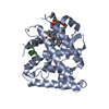
Mass: 766.124 Da / Num. of mol.: 1 / Source method: obtained synthetically / Formula: C42H88NO8P / Comment: SPPC, phospholipid*YM
#4: Water ChemComp-HOH / water / Water


Mass: 18.015 Da / Num. of mol.: 123 / Source method: isolated from a natural source / Formula: H2O

-
Experimental details

-
Experiment

ExperimentMethod: X-RAY DIFFRACTION / Number of used crystals: 1

-
Sample preparation

CrystalDensity Matthews: 2.21 Å3/Da / Density % sol: 44.34 %
Crystal growTemperature: 298 K / Method: vapor diffusion, hanging drop / pH: 6.5
Details: 0.1 M sodium cacodylate, 20% PEG 3350, 0.2 M sodium acetate, 2 mM TCEP, 10% xylitol, pH 6.5, VAPOR DIFFUSION, HANGING DROP, temperature 298K

-
Data collection

DiffractionMean temperature: 100 K
Diffraction sourceSource: SYNCHROTRON / Site: ALS / Beamline: 8.3.1 / Wavelength: 1.1 Å
DetectorType: ADSC QUANTUM 315 / Detector: CCD / Date: Jun 25, 2005 / Details: mirrors
RadiationMonochromator: KHOZU Double flat crystal Si(111) / Protocol: SINGLE WAVELENGTH / Monochromatic (M) / Laue (L): M / Scattering type: x-ray
Radiation wavelengthWavelength: 1.1 Å / Relative weight: 1
ReflectionResolution: 2.2→25 Å / Num. all: 13803 / Num. obs: 13232 / % possible obs: 95.86 % / Observed criterion σ(F): 0 / Observed criterion σ(I): 0 / Redundancy: 3.7 % / Biso Wilson estimate: 26.5 Å2 / Rmerge(I) obs: 0.07 / Rsym value: 0.07 / Net I/σ(I): 15.1
Reflection shellResolution: 2.2→2.28 Å / Redundancy: 2.7 % / Rmerge(I) obs: 0.24 / Mean I/σ(I) obs: 3.3 / Rsym value: 0.24 / % possible all: 89.5

-
Processing

Software
NameVersionClassification
PHENIX(phenix.refine)refinement
CNSrefinement
ADSCQuantumdata collection
DENZOdata reduction
SCALEPACKdata scaling
CNSphasing
RefinementMethod to determine structure: MOLECULAR REPLACEMENT
Starting model: PDb entry 1YMT
Resolution: 2.2→23.76 Å / SU ML: 0.32 / σ(F): 0.06 / Stereochemistry target values: ML
RfactorNum. reflection% reflection
Rfree0.241 399 3.02 %
Rwork0.18 --
obs0.182 13232 95.9 %
all-13803 -
Solvent computationShrinkage radii: 0.9 Å / VDW probe radii: 1.11 Å / Solvent model: FLAT BULK SOLVENT MODEL / Bsol: 47.77 Å2 / ksol: 0.34 e/Å3
Refinement stepCycle: LAST / Resolution: 2.2→23.76 Å
ProteinNucleic acidLigandSolventTotal
Num. atoms1996 0 52 123 2171
Refine LS restraints
Refine-IDTypeDev ideal
X-RAY DIFFRACTIONf_bond_d0.008
X-RAY DIFFRACTIONf_angle_d1.006
X-RAY DIFFRACTIONf_dihedral_angle_d
X-RAY DIFFRACTIONf_chiral_restr
X-RAY DIFFRACTIONf_plane_restr
LS refinement shell
Resolution (Å)Rfactor RfreeNum. reflection RfreeRfactor RworkNum. reflection RworkRefine-ID% reflection obs (%)
2.2-2.51820.26641300.21583989X-RAY DIFFRACTION92
2.5182-3.17150.26171200.19384303X-RAY DIFFRACTION97
3.1715-23.76380.22181490.16064541X-RAY DIFFRACTION99
Refinement TLS params.

Method: refined / Refine-ID: X-RAY DIFFRACTION

IDL112)L122)L132)L222)L232)L332)S11 (Å °)S12 (Å °)S13 (Å °)S21 (Å °)S22 (Å °)S23 (Å °)S31 (Å °)S32 (Å °)S33 (Å °)T112)T122)T132)T222)T232)T332)Origin x (Å)Origin y (Å)Origin z (Å)
10.92790.0403-0.05321.59210.32221.0437-0.0001-0.0051-0.0471-0.13370.04420.1078-0.0475-0.0151-0.03880.03260.0037-0.00680.03250.02390.0348-76.298940.7911-25.0635
20.02810.03280.0790.38120.10110.30920.02070.0954-0.0768-0.2550.01210.1732-0.03430.09520.02970.4318-0.0579-0.12520.323-0.21360.2899-84.573635.9774-42.0542
3-0.25940.17990.04310.17590.03270.06520.15650.1275-0.07350.03060.2819-0.1042-0.0386-0.1714-0.36170.15020.02980.0320.32120.050.2854-61.552236.7247-28.7616
Refinement TLS group
IDRefine-IDRefine TLS-IDSelection details
1X-RAY DIFFRACTION1CHAIN A
2X-RAY DIFFRACTION2CHAIN B
3X-RAY DIFFRACTION3CHAIN C

+
About Yorodumi

-
News

-
Feb 9, 2022. New format data for meta-information of EMDB entries

New format data for meta-information of EMDB entries

  • Version 3 of the EMDB header file is now the official format.
  • The previous official version 1.9 will be removed from the archive.

Related info.:EMDB header

External links:wwPDB to switch to version 3 of the EMDB data model

-
Aug 12, 2020. Covid-19 info

Covid-19 info

URL: https://pdbj.org/emnavi/covid19.php

New page: Covid-19 featured information page in EM Navigator.

Related info.:Covid-19 info / Mar 5, 2020. Novel coronavirus structure data

+
Mar 5, 2020. Novel coronavirus structure data

Novel coronavirus structure data

Related info.:Yorodumi Speices / Aug 12, 2020. Covid-19 info

External links:COVID-19 featured content - PDBj / Molecule of the Month (242):Coronavirus Proteases

+
Jan 31, 2019. EMDB accession codes are about to change! (news from PDBe EMDB page)

EMDB accession codes are about to change! (news from PDBe EMDB page)

  • The allocation of 4 digits for EMDB accession codes will soon come to an end. Whilst these codes will remain in use, new EMDB accession codes will include an additional digit and will expand incrementally as the available range of codes is exhausted. The current 4-digit format prefixed with “EMD-” (i.e. EMD-XXXX) will advance to a 5-digit format (i.e. EMD-XXXXX), and so on. It is currently estimated that the 4-digit codes will be depleted around Spring 2019, at which point the 5-digit format will come into force.
  • The EM Navigator/Yorodumi systems omit the EMD- prefix.

Related info.:Q: What is EMD? / ID/Accession-code notation in Yorodumi/EM Navigator

External links:EMDB Accession Codes are Changing Soon! / Contact to PDBj

+
Jul 12, 2017. Major update of PDB

Major update of PDB

  • wwPDB released updated PDB data conforming to the new PDBx/mmCIF dictionary.
  • This is a major update changing the version number from 4 to 5, and with Remediation, in which all the entries are updated.
  • In this update, many items about electron microscopy experimental information are reorganized (e.g. em_software).
  • Now, EM Navigator and Yorodumi are based on the updated data.

External links:wwPDB Remediation / Enriched Model Files Conforming to OneDep Data Standards Now Available in the PDB FTP Archive

-
Yorodumi

Thousand views of thousand structures

  • Yorodumi is a browser for structure data from EMDB, PDB, SASBDB, etc.
  • This page is also the successor to EM Navigator detail page, and also detail information page/front-end page for Omokage search.
  • The word "yorodu" (or yorozu) is an old Japanese word meaning "ten thousand". "mi" (miru) is to see.

Related info.:EMDB / PDB / SASBDB / Comparison of 3 databanks / Yorodumi Search / Aug 31, 2016. New EM Navigator & Yorodumi / Yorodumi Papers / Jmol/JSmol / Function and homology information / Changes in new EM Navigator and Yorodumi

Read more