[English] 日本語
Yorodumi
- PDB-3f5h: Crystal structure of fused docking domains from PikAIII and PikAI... -

+
Open data


ID or keywords:

Loading...

-
Basic information

Entry
Database: PDB / ID: 3f5h
TitleCrystal structure of fused docking domains from PikAIII and PikAIV of the pikromycin polyketide synthase
ComponentsType I polyketide synthase PikAIII, Type I polyketide synthase PikAIV fusion protein
KeywordsPROTEIN BINDING / docking domain / polyketide synthase / pikromycin / H2-T2
Function / homology
Function and homology information


10-deoxymethynolide synthase / narbonolide synthase / macrolide biosynthetic process / DIM/DIP cell wall layer assembly / fatty acid synthase activity / acyltransferase activity, transferring groups other than amino-acyl groups / phosphopantetheine binding / 3-oxoacyl-[acyl-carrier-protein] synthase activity / fatty acid biosynthetic process / identical protein binding ...10-deoxymethynolide synthase / narbonolide synthase / macrolide biosynthetic process / DIM/DIP cell wall layer assembly / fatty acid synthase activity / acyltransferase activity, transferring groups other than amino-acyl groups / phosphopantetheine binding / 3-oxoacyl-[acyl-carrier-protein] synthase activity / fatty acid biosynthetic process / identical protein binding / plasma membrane / cytoplasm
Similarity search - Function
Polyketide synthase, docking domain superfmaily / Polyketide synthase, thioesterase domain / Thioesterase / Polyketide synthase, docking domain / Erythronolide synthase docking domain / PKS_PP_betabranch / Thioesterase / Thioesterase domain / Polyketide synthase, ketoreductase domain / KR domain ...Polyketide synthase, docking domain superfmaily / Polyketide synthase, thioesterase domain / Thioesterase / Polyketide synthase, docking domain / Erythronolide synthase docking domain / PKS_PP_betabranch / Thioesterase / Thioesterase domain / Polyketide synthase, ketoreductase domain / KR domain / Malonyl-CoA ACP transacylase, ACP-binding / Polyketide synthase, C-terminal extension / Ketoacyl-synthetase C-terminal extension / PKS_KR / Acyl transferase domain superfamily / Acyl transferase / Acyl transferase domain / Acyl transferase domain in polyketide synthase (PKS) enzymes. / Acyl transferase/acyl hydrolase/lysophospholipase / Ketosynthase family 3 (KS3) domain profile. / Polyketide synthase, phosphopantetheine-binding domain / Phosphopantetheine attachment site / Beta-ketoacyl synthase / Beta-ketoacyl synthase, active site / Ketosynthase family 3 (KS3) active site signature. / Polyketide synthase, beta-ketoacyl synthase domain / Beta-ketoacyl synthase, N-terminal / Beta-ketoacyl synthase, C-terminal / Beta-ketoacyl synthase, N-terminal domain / Beta-ketoacyl synthase, C-terminal domain / Thiolase-like / Phosphopantetheine attachment site / Phosphopantetheine attachment site. / Phosphopantetheine attachment site / ACP-like superfamily / Carrier protein (CP) domain profile. / Phosphopantetheine binding ACP domain / Alpha/Beta hydrolase fold / NAD(P)-binding domain superfamily
Similarity search - Domain/homology
Narbonolide/10-deoxymethynolide synthase PikA4, module 6 / Narbonolide/10-deoxymethynolide synthase PikA3, module 5
Similarity search - Component
Biological speciesStreptomyces venezuelae (bacteria)
MethodX-RAY DIFFRACTION / SYNCHROTRON / SAD / Resolution: 1.75 Å
AuthorsBuchholz, T.J. / Geders, T.W. / Bartley, F.E. / Reynolds, K.A. / Smith, J.L. / Sherman, D.H.
CitationJournal: Acs Chem.Biol. / Year: 2009
Title: Structural basis for binding specificity between subclasses of modular polyketide synthase docking domains.
Authors: Buchholz, T.J. / Geders, T.W. / Bartley, F.E. / Reynolds, K.A. / Smith, J.L. / Sherman, D.H.
History
DepositionNov 3, 2008Deposition site: RCSB / Processing site: RCSB
Revision 1.0Jan 27, 2009Provider: repository / Type: Initial release
Revision 1.1Jul 13, 2011Group: Version format compliance
Revision 1.2Aug 2, 2017Group: Refinement description / Source and taxonomy / Category: entity_src_gen / software
Revision 1.3Oct 25, 2017Group: Refinement description / Category: software
Revision 1.4Dec 27, 2023Group: Data collection / Database references / Derived calculations
Category: chem_comp_atom / chem_comp_bond ...chem_comp_atom / chem_comp_bond / database_2 / pdbx_struct_conn_angle / struct_conn / struct_ref_seq_dif
Item: _database_2.pdbx_DOI / _database_2.pdbx_database_accession ..._database_2.pdbx_DOI / _database_2.pdbx_database_accession / _pdbx_struct_conn_angle.ptnr1_auth_comp_id / _pdbx_struct_conn_angle.ptnr1_auth_seq_id / _pdbx_struct_conn_angle.ptnr1_label_asym_id / _pdbx_struct_conn_angle.ptnr1_label_atom_id / _pdbx_struct_conn_angle.ptnr1_label_comp_id / _pdbx_struct_conn_angle.ptnr1_label_seq_id / _pdbx_struct_conn_angle.ptnr3_auth_comp_id / _pdbx_struct_conn_angle.ptnr3_auth_seq_id / _pdbx_struct_conn_angle.ptnr3_label_asym_id / _pdbx_struct_conn_angle.ptnr3_label_atom_id / _pdbx_struct_conn_angle.ptnr3_label_comp_id / _pdbx_struct_conn_angle.ptnr3_label_seq_id / _pdbx_struct_conn_angle.value / _struct_conn.pdbx_dist_value / _struct_conn.ptnr1_auth_comp_id / _struct_conn.ptnr1_auth_seq_id / _struct_conn.ptnr1_label_asym_id / _struct_conn.ptnr1_label_atom_id / _struct_conn.ptnr1_label_comp_id / _struct_conn.ptnr1_label_seq_id / _struct_conn.ptnr2_auth_comp_id / _struct_conn.ptnr2_auth_seq_id / _struct_conn.ptnr2_label_asym_id / _struct_conn.ptnr2_label_atom_id / _struct_conn.ptnr2_label_comp_id / _struct_ref_seq_dif.details

-
Structure visualization

Structure viewerMolecule:
MolmilJmol/JSmol

Downloads & links

-
Assembly

Deposited unit
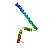
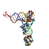
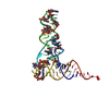
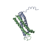
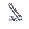
A: Type I polyketide synthase PikAIII, Type I polyketide synthase PikAIV fusion protein
B: Type I polyketide synthase PikAIII, Type I polyketide synthase PikAIV fusion protein
hetero molecules


Theoretical massNumber of molelcules
Total (without water)15,2923
Polymers15,2692
Non-polymers231
Water2,720151
1


  • Idetical with deposited unit
  • defined by software
TypeNameSymmetry operationNumber
identity operation1_555x,y,z1
Buried area1830 Å2
ΔGint-20 kcal/mol
Surface area9320 Å2
MethodPISA
Unit cell
Length a, b, c (Å)59.016, 117.944, 41.773
Angle α, β, γ (deg.)90.00, 90.00, 90.00
Int Tables number20
Space group name H-MC2221
DetailsTHE CONTENTS OF THE ASYMMETRIC UNIT IS AN UNNATURAL FUSION OF THE C-TERMINUS OF ONE PROTEIN TO THE N-TERMINUS OF A SECOND PROTEIN. THE BIOLOGICAL ASSEMBLY IS FORMED PARTIALLY THROUGH CRYSTALLOGRAPHIC SYMMETRY. THE RELEVANT BIOLOGICAL ASSEMBLY IS FORMED BY APPLYING THE FOLLOWING TRANSFORMATIONS: RESIDUES 1-34 OF CHAIN A AND RESIDUES 1-37 OF CHAIN B:[X, Y, Z]. RESIDUES 1543-1562 OF CHAIN A:[-X+3/2, -Y+1/2, Z-1/2]. RESIDUES 1543-1562 OF CHAIN B:[-X+3/2, -Y+1/2, Z+1/2].

-
Components

#1: Protein Type I polyketide synthase PikAIII, Type I polyketide synthase PikAIV fusion protein


Mass: 7634.411 Da / Num. of mol.: 2
Fragment: C-terminal domain of PikAIII fused to N-terminal domain of PikAIV
Source method: isolated from a genetically manipulated source
Source: (gene. exp.) Streptomyces venezuelae (bacteria) / Strain: ATCC15439 / Gene: pIKAIII, pIKAIV / Plasmid: pMCSG7 / Production host: Escherichia coli (E. coli) / Strain (production host): BL21(DE3) / References: UniProt: Q9ZGI3, UniProt: Q9ZGI2
#2: Chemical ChemComp-NA / SODIUM ION


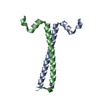
Mass: 22.990 Da / Num. of mol.: 1 / Source method: obtained synthetically / Formula: Na
#3: Water ChemComp-HOH / water


Mass: 18.015 Da / Num. of mol.: 151 / Source method: isolated from a natural source / Formula: H2O

-
Experimental details

-
Experiment

ExperimentMethod: X-RAY DIFFRACTION / Number of used crystals: 1

-
Sample preparation

CrystalDensity Matthews: 2.38 Å3/Da / Density % sol: 48.33 %
Crystal growTemperature: 277 K / Method: vapor diffusion, hanging drop / pH: 5
Details: 20 mM HEPES pH 7.8, 150 mM NaCl, 55% 2-methyl-2,4-pentanediol, 200 mM sodium acetate pH 5.0, VAPOR DIFFUSION, HANGING DROP, temperature 277K

-
Data collection

Diffraction
IDMean temperature (K)Crystal-ID
11001
21001
Diffraction source
SourceSiteBeamlineIDWavelength (Å)
SYNCHROTRONAPS 23-ID-D10.97934
SYNCHROTRONAPS 23-ID-B20.9794
Detector
TypeIDDetectorDateDetails
MARMOSAIC 300 mm CCD1CCDFeb 10, 2008K-B PAIR OF BIOMORPH MIRRORS FOR VERTICAL AND HORIZONTAL FOCUSING
MARMOSAIC 300 mm CCD2CCDApr 21, 2008K-B PAIR OF BIOMORPH MIRRORS FOR VERTICAL AND HORIZONTAL FOCUSING
Radiation
IDMonochromatorProtocolMonochromatic (M) / Laue (L)Scattering typeWavelength-ID
1DOUBLE CRYSTAL MONOCHROMATORSINGLE WAVELENGTHMx-ray1
2DOUBLE CRYSTAL MONOCHROMATORSINGLE WAVELENGTHMx-ray1
Radiation wavelength
IDWavelength (Å)Relative weight
10.979341
20.97941
ReflectionRedundancy: 5.1 % / Av σ(I) over netI: 15.59 / Number: 36536 / Rmerge(I) obs: 0.123 / Χ2: 0.98 / D res high: 2.8 Å / D res low: 50 Å / Num. obs: 7102 / % possible obs: 100
Diffraction reflection shell
Highest resolution (Å)Lowest resolution (Å)% possible obs (%)IDRmerge(I) obsChi squaredRedundancy
6.035099.910.0910.9485.1
4.796.0310010.0880.9695.1
4.184.7910010.0971.0195.2
3.84.1810010.0970.9765.2
3.533.810010.1111.0475.2
3.323.5310010.1320.9475.1
3.153.3210010.1531.0365.2
3.023.1510010.2051.0025.2
2.93.0210010.2250.9235.1
2.82.910010.2970.9345.1
ReflectionResolution: 1.75→50 Å / Num. all: 15084 / Num. obs: 14954 / % possible obs: 99.2 % / Observed criterion σ(F): 0 / Observed criterion σ(I): 0 / Redundancy: 3.6 % / Biso Wilson estimate: 20.3 Å2 / Rmerge(I) obs: 0.069 / Rsym value: 0.069 / Χ2: 0.996 / Net I/σ(I): 15.586
Reflection shellResolution: 1.75→1.81 Å / Redundancy: 3.1 % / Rmerge(I) obs: 0.507 / Mean I/σ(I) obs: 2.1 / Num. unique all: 1471 / Rsym value: 0.507 / Χ2: 0.993 / % possible all: 98

-
Phasing

PhasingMethod: SAD
Phasing dmFOM : 0.36 / FOM acentric: 0.37 / FOM centric: 0.33 / Reflection: 3089 / Reflection acentric: 2531 / Reflection centric: 558
Phasing dm shell
Resolution (Å)FOM FOM acentricFOM centricReflectionReflection acentricReflection centric
8.6-34.1970.490.550.421538865
5.4-8.60.340.350.29423316107
4.3-5.40.430.450.37528427101
3.8-4.30.460.470.453043496
3.2-3.80.330.340.26904783121
3-3.20.240.230.355148368

-
Processing

Software
NameVersionClassificationNB
DENZOdata reduction
SCALEPACKdata scaling
SOLVE2.13phasing
RESOLVE2.13phasing
REFMACrefinement
PDB_EXTRACT3.006data extraction
HKL-2000data collection
RefinementMethod to determine structure: SAD / Resolution: 1.75→32.76 Å / Cor.coef. Fo:Fc: 0.955 / Cor.coef. Fo:Fc free: 0.932 / WRfactor Rfree: 0.25 / WRfactor Rwork: 0.206 / Occupancy max: 1 / Occupancy min: 0.5 / FOM work R set: 0.801 / SU B: 2.945 / SU ML: 0.091 / SU R Cruickshank DPI: 0.118 / SU Rfree: 0.123 / Isotropic thermal model: ISOTROPIC / Cross valid method: THROUGHOUT / σ(F): 0 / σ(I): 0 / ESU R: 0.118 / ESU R Free: 0.123 / Stereochemistry target values: MAXIMUM LIKELIHOOD / Details: HYDROGENS HAVE BEEN ADDED IN THE RIDING POSITIONS
RfactorNum. reflection% reflectionSelection details
Rfree0.25 750 5 %RANDOM
Rwork0.201 ---
all0.203 15079 --
obs0.203 14942 99.09 %-
Solvent computationIon probe radii: 0.8 Å / Shrinkage radii: 0.8 Å / VDW probe radii: 1.2 Å / Solvent model: BABINET MODEL WITH MASK
Displacement parametersBiso max: 63.09 Å2 / Biso mean: 26.546 Å2 / Biso min: 12.69 Å2
Baniso -1Baniso -2Baniso -3
1-3.52 Å20 Å20 Å2
2---1.88 Å20 Å2
3----1.65 Å2
Refinement stepCycle: LAST / Resolution: 1.75→32.76 Å
ProteinNucleic acidLigandSolventTotal
Num. atoms919 0 1 151 1071
Refine LS restraints
Refine-IDTypeDev idealDev ideal targetNumber
X-RAY DIFFRACTIONr_bond_refined_d0.0110.021926
X-RAY DIFFRACTIONr_angle_refined_deg1.2162.0011244
X-RAY DIFFRACTIONr_dihedral_angle_1_deg3.9585120
X-RAY DIFFRACTIONr_dihedral_angle_2_deg34.24122.8353
X-RAY DIFFRACTIONr_dihedral_angle_3_deg12.76515198
X-RAY DIFFRACTIONr_dihedral_angle_4_deg16.8771519
X-RAY DIFFRACTIONr_chiral_restr0.0710.2139
X-RAY DIFFRACTIONr_gen_planes_refined0.0050.021707
X-RAY DIFFRACTIONr_mcbond_it0.8021.5573
X-RAY DIFFRACTIONr_mcangle_it1.6522921
X-RAY DIFFRACTIONr_scbond_it3.2113353
X-RAY DIFFRACTIONr_scangle_it5.7594.5317
LS refinement shellResolution: 1.752→1.797 Å / Total num. of bins used: 20
RfactorNum. reflection% reflection
Rfree0.258 59 -
Rwork0.281 982 -
all-1041 -
obs--96.39 %

+
About Yorodumi

-
News

-
Feb 9, 2022. New format data for meta-information of EMDB entries

New format data for meta-information of EMDB entries

  • Version 3 of the EMDB header file is now the official format.
  • The previous official version 1.9 will be removed from the archive.

Related info.:EMDB header

External links:wwPDB to switch to version 3 of the EMDB data model

-
Aug 12, 2020. Covid-19 info

Covid-19 info

URL: https://pdbjlvh1.pdbj.org/emnavi/covid19.php

New page: Covid-19 featured information page in EM Navigator.

Related info.:Covid-19 info / Mar 5, 2020. Novel coronavirus structure data

+
Mar 5, 2020. Novel coronavirus structure data

Novel coronavirus structure data

Related info.:Yorodumi Speices / Aug 12, 2020. Covid-19 info

External links:COVID-19 featured content - PDBj / Molecule of the Month (242):Coronavirus Proteases

+
Jan 31, 2019. EMDB accession codes are about to change! (news from PDBe EMDB page)

EMDB accession codes are about to change! (news from PDBe EMDB page)

  • The allocation of 4 digits for EMDB accession codes will soon come to an end. Whilst these codes will remain in use, new EMDB accession codes will include an additional digit and will expand incrementally as the available range of codes is exhausted. The current 4-digit format prefixed with “EMD-” (i.e. EMD-XXXX) will advance to a 5-digit format (i.e. EMD-XXXXX), and so on. It is currently estimated that the 4-digit codes will be depleted around Spring 2019, at which point the 5-digit format will come into force.
  • The EM Navigator/Yorodumi systems omit the EMD- prefix.

Related info.:Q: What is EMD? / ID/Accession-code notation in Yorodumi/EM Navigator

External links:EMDB Accession Codes are Changing Soon! / Contact to PDBj

+
Jul 12, 2017. Major update of PDB

Major update of PDB

  • wwPDB released updated PDB data conforming to the new PDBx/mmCIF dictionary.
  • This is a major update changing the version number from 4 to 5, and with Remediation, in which all the entries are updated.
  • In this update, many items about electron microscopy experimental information are reorganized (e.g. em_software).
  • Now, EM Navigator and Yorodumi are based on the updated data.

External links:wwPDB Remediation / Enriched Model Files Conforming to OneDep Data Standards Now Available in the PDB FTP Archive

-
Yorodumi

Thousand views of thousand structures

  • Yorodumi is a browser for structure data from EMDB, PDB, SASBDB, etc.
  • This page is also the successor to EM Navigator detail page, and also detail information page/front-end page for Omokage search.
  • The word "yorodu" (or yorozu) is an old Japanese word meaning "ten thousand". "mi" (miru) is to see.

Related info.:EMDB / PDB / SASBDB / Comparison of 3 databanks / Yorodumi Search / Aug 31, 2016. New EM Navigator & Yorodumi / Yorodumi Papers / Jmol/JSmol / Function and homology information / Changes in new EM Navigator and Yorodumi

Read more