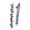
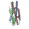
[English] 日本語
Yorodumi
- PDB-3f4y: HIV gp41 six-helix bundle containing a mutant CHR alpha-peptide s... -

+
Open data


ID or keywords:

Loading...

-
Basic information

Entry
Database: PDB / ID: 3f4y
TitleHIV gp41 six-helix bundle containing a mutant CHR alpha-peptide sequence
Components
  • Envelope glycoprotein gp160
  • Mutant peptide derived from HIV gp41 CHR domain
KeywordsVIRAL PROTEIN / helix bundle / AIDS / Apoptosis / Cell membrane / Cleavage on pair of basic residues / Coiled coil / Envelope protein / Fusion protein / Glycoprotein / Host-virus interaction / Lipoprotein / Membrane / Palmitate / Transmembrane / Viral immunoevasion / Virion
Function / homology
Function and homology information


Dectin-2 family / positive regulation of plasma membrane raft polarization / positive regulation of receptor clustering / positive regulation of establishment of T cell polarity / virus-mediated perturbation of host defense response / host cell endosome membrane / clathrin-dependent endocytosis of virus by host cell / viral protein processing / fusion of virus membrane with host plasma membrane / fusion of virus membrane with host endosome membrane ...Dectin-2 family / positive regulation of plasma membrane raft polarization / positive regulation of receptor clustering / positive regulation of establishment of T cell polarity / virus-mediated perturbation of host defense response / host cell endosome membrane / clathrin-dependent endocytosis of virus by host cell / viral protein processing / fusion of virus membrane with host plasma membrane / fusion of virus membrane with host endosome membrane / viral envelope / virion attachment to host cell / host cell plasma membrane / virion membrane / structural molecule activity / membrane
Similarity search - Function
Envelope glycoprotein Gp160 / Retroviral envelope protein / Retroviral envelope protein GP41-like / Gp120 core superfamily / Envelope glycoprotein GP120 / Human immunodeficiency virus 1, envelope glycoprotein Gp120
Similarity search - Domain/homology
Envelope glycoprotein gp160
Similarity search - Component
MethodX-RAY DIFFRACTION / Resolution: 2 Å
AuthorsHorne, W.S. / Gellman, S.H.
CitationJournal: Proc.Natl.Acad.Sci.USA / Year: 2009
Title: Structural and biological mimicry of protein surface recognition by alpha/beta-peptide foldamers
Authors: Horne, W.S. / Johnson, L.M. / Ketas, T.J. / Klasse, P.J. / Lu, M. / Moore, J.P. / Gellman, S.H.
History
DepositionNov 3, 2008Deposition site: RCSB / Processing site: RCSB
Revision 1.0Sep 15, 2009Provider: repository / Type: Initial release
Revision 1.1Jul 13, 2011Group: Version format compliance
Revision 1.2Dec 27, 2023Group: Data collection / Database references / Derived calculations
Category: chem_comp_atom / chem_comp_bond ...chem_comp_atom / chem_comp_bond / database_2 / struct_conn / struct_ref_seq
Item: _database_2.pdbx_DOI / _database_2.pdbx_database_accession ..._database_2.pdbx_DOI / _database_2.pdbx_database_accession / _struct_conn.pdbx_leaving_atom_flag / _struct_ref_seq.db_align_beg / _struct_ref_seq.db_align_end

-
Structure visualization

Structure viewerMolecule:
MolmilJmol/JSmol

Downloads & links

-
Assembly

Deposited unit
A: Envelope glycoprotein gp160
B: Envelope glycoprotein gp160
C: Envelope glycoprotein gp160
D: Mutant peptide derived from HIV gp41 CHR domain
E: Mutant peptide derived from HIV gp41 CHR domain
F: Mutant peptide derived from HIV gp41 CHR domain


Theoretical massNumber of molelcules
Total (without water)25,7876
Polymers25,7876
Non-polymers00
Water2,738152
1


  • Idetical with deposited unit
  • defined by author&software
TypeNameSymmetry operationNumber
identity operation1_555x,y,z1
Buried area11770 Å2
ΔGint-101 kcal/mol
Surface area11670 Å2
MethodPISA
Unit cell
Length a, b, c (Å)37.611, 179.038, 33.075
Angle α, β, γ (deg.)90.000, 90.000, 90.000
Int Tables number18
Space group name H-MP21212
Components on special symmetry positions
IDModelComponents
11B-151-

HOH

-
Components

#1: Protein/peptide Envelope glycoprotein gp160 / Env polyprotein / Surface protein / SU / Glycoprotein 120 / gp120 / Transmembrane protein / TM / ...Env polyprotein / Surface protein / SU / Glycoprotein 120 / gp120 / Transmembrane protein / TM / Glycoprotein 41 / gp41


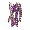
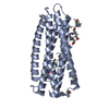
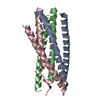
Mass: 4150.851 Da / Num. of mol.: 3 / Fragment: HIV gp41 NHR domain / Source method: obtained synthetically
Details: The peptide is chemically synthesized. It occurs naturally in HIV.
References: UniProt: P04580
#2: Protein/peptide Mutant peptide derived from HIV gp41 CHR domain


Mass: 4444.915 Da / Num. of mol.: 3 / Fragment: HIV gp41 CHR domain mutant
Mutation: M1T, M4E, E5A, E9A, N11A, N12E, T14A, S15A, L16R, H18E, S19A, N21E, Q33A, E34A, L36R
Source method: obtained synthetically
Details: The peptide is chemically synthesized. It is a sequence mutant to a sequence that occurs naturally in HIV.
#3: Water ChemComp-HOH / water / Water


Mass: 18.015 Da / Num. of mol.: 152 / Source method: isolated from a natural source / Formula: H2O

-
Experimental details

-
Experiment

ExperimentMethod: X-RAY DIFFRACTION / Number of used crystals: 1

-
Sample preparation

CrystalDensity Matthews: 2.16 Å3/Da / Density % sol: 43.03 %
Crystal growTemperature: 298 K / Method: vapor diffusion, hanging drop / pH: 8.5
Details: 0.1 M Tris pH 8.5, 1 M ammonium phosphate monobasic, VAPOR DIFFUSION, HANGING DROP, temperature 298K

-
Data collection

DiffractionMean temperature: 100 K
Diffraction sourceSource: ROTATING ANODE / Type: BRUKER AXS MICROSTAR / Wavelength: 1.5418 Å
DetectorType: BRUKER SMART 6000 / Detector: CCD / Date: Nov 5, 2007 / Details: confocal mirrors
RadiationMonochromator: gobel mirrors / Protocol: SINGLE WAVELENGTH / Monochromatic (M) / Laue (L): M / Scattering type: x-ray
Radiation wavelengthWavelength: 1.5418 Å / Relative weight: 1
ReflectionResolution: 2→44.8 Å / Num. all: 15956 / Num. obs: 15938 / % possible obs: 99.9 % / Redundancy: 8.6 % / Rsym value: 0.05 / Net I/σ(I): 28
Reflection shellResolution: 2→2.1 Å / Redundancy: 3.6 % / Mean I/σ(I) obs: 4.7 / Rsym value: 0.262 / % possible all: 100

-
Processing

Software
NameVersionClassificationNB
REFMAC5.3.0037refinement
PDB_EXTRACT3.006data extraction
PROTEUM PLUSPLUSdata collection
PROTEUM PLUSPLUSdata reduction
PROTEUM PLUSPLUSdata scaling
PHASERphasing
RefinementResolution: 2→25 Å / Cor.coef. Fo:Fc: 0.933 / Cor.coef. Fo:Fc free: 0.901 / Occupancy max: 1 / Occupancy min: 0 / SU B: 5.203 / SU ML: 0.147 / Cross valid method: THROUGHOUT / σ(F): 0 / ESU R: 0.227 / ESU R Free: 0.193 / Stereochemistry target values: MAXIMUM LIKELIHOOD / Details: HYDROGENS HAVE BEEN ADDED IN THE RIDING POSITIONS
RfactorNum. reflection% reflectionSelection details
Rfree0.26 758 4.8 %RANDOM
Rwork0.209 ---
obs0.212 15881 99.96 %-
Solvent computationIon probe radii: 0.8 Å / Shrinkage radii: 0.8 Å / VDW probe radii: 1.4 Å / Solvent model: MASK
Displacement parametersBiso max: 62.36 Å2 / Biso mean: 24.219 Å2 / Biso min: 2 Å2
Baniso -1Baniso -2Baniso -3
1--0.06 Å20 Å20 Å2
2---0.55 Å20 Å2
3---0.61 Å2
Refinement stepCycle: LAST / Resolution: 2→25 Å
ProteinNucleic acidLigandSolventTotal
Num. atoms3769 0 0 456 4225
Refine LS restraints
Refine-IDTypeDev idealDev ideal targetNumber
X-RAY DIFFRACTIONr_bond_refined_d0.0130.0221887
X-RAY DIFFRACTIONr_bond_other_d0.0010.021268
X-RAY DIFFRACTIONr_angle_refined_deg1.1461.9352562
X-RAY DIFFRACTIONr_angle_other_deg0.933084
X-RAY DIFFRACTIONr_dihedral_angle_1_deg4.7295227
X-RAY DIFFRACTIONr_dihedral_angle_2_deg34.91125.094106
X-RAY DIFFRACTIONr_dihedral_angle_3_deg18.30915351
X-RAY DIFFRACTIONr_dihedral_angle_4_deg16.5191518
X-RAY DIFFRACTIONr_chiral_restr0.0660.2289
X-RAY DIFFRACTIONr_gen_planes_refined0.0040.022094
X-RAY DIFFRACTIONr_gen_planes_other0.0010.02371
X-RAY DIFFRACTIONr_nbd_refined0.2050.2506
X-RAY DIFFRACTIONr_nbd_other0.1760.21354
X-RAY DIFFRACTIONr_nbtor_refined0.170.2890
X-RAY DIFFRACTIONr_nbtor_other0.090.21035
X-RAY DIFFRACTIONr_xyhbond_nbd_refined0.210.2104
X-RAY DIFFRACTIONr_symmetry_vdw_refined0.1580.230
X-RAY DIFFRACTIONr_symmetry_vdw_other0.3120.276
X-RAY DIFFRACTIONr_symmetry_hbond_refined0.2760.211
X-RAY DIFFRACTIONr_mcbond_it1.5251.51536
X-RAY DIFFRACTIONr_mcbond_other0.1451.5468
X-RAY DIFFRACTIONr_mcangle_it1.24721809
X-RAY DIFFRACTIONr_scbond_it2.7923998
X-RAY DIFFRACTIONr_scangle_it3.1974.5747
LS refinement shellResolution: 2→2.052 Å / Total num. of bins used: 20
RfactorNum. reflection% reflection
Rfree0.398 49 -
Rwork0.248 1079 -
all-1128 -
obs--99.91 %

+
About Yorodumi

-
News

-
Feb 9, 2022. New format data for meta-information of EMDB entries

New format data for meta-information of EMDB entries

  • Version 3 of the EMDB header file is now the official format.
  • The previous official version 1.9 will be removed from the archive.

Related info.:EMDB header

External links:wwPDB to switch to version 3 of the EMDB data model

-
Aug 12, 2020. Covid-19 info

Covid-19 info

URL: https://pdbj.org/emnavi/covid19.php

New page: Covid-19 featured information page in EM Navigator.

Related info.:Covid-19 info / Mar 5, 2020. Novel coronavirus structure data

+
Mar 5, 2020. Novel coronavirus structure data

Novel coronavirus structure data

Related info.:Yorodumi Speices / Aug 12, 2020. Covid-19 info

External links:COVID-19 featured content - PDBj / Molecule of the Month (242):Coronavirus Proteases

+
Jan 31, 2019. EMDB accession codes are about to change! (news from PDBe EMDB page)

EMDB accession codes are about to change! (news from PDBe EMDB page)

  • The allocation of 4 digits for EMDB accession codes will soon come to an end. Whilst these codes will remain in use, new EMDB accession codes will include an additional digit and will expand incrementally as the available range of codes is exhausted. The current 4-digit format prefixed with “EMD-” (i.e. EMD-XXXX) will advance to a 5-digit format (i.e. EMD-XXXXX), and so on. It is currently estimated that the 4-digit codes will be depleted around Spring 2019, at which point the 5-digit format will come into force.
  • The EM Navigator/Yorodumi systems omit the EMD- prefix.

Related info.:Q: What is EMD? / ID/Accession-code notation in Yorodumi/EM Navigator

External links:EMDB Accession Codes are Changing Soon! / Contact to PDBj

+
Jul 12, 2017. Major update of PDB

Major update of PDB

  • wwPDB released updated PDB data conforming to the new PDBx/mmCIF dictionary.
  • This is a major update changing the version number from 4 to 5, and with Remediation, in which all the entries are updated.
  • In this update, many items about electron microscopy experimental information are reorganized (e.g. em_software).
  • Now, EM Navigator and Yorodumi are based on the updated data.

External links:wwPDB Remediation / Enriched Model Files Conforming to OneDep Data Standards Now Available in the PDB FTP Archive

-
Yorodumi

Thousand views of thousand structures

  • Yorodumi is a browser for structure data from EMDB, PDB, SASBDB, etc.
  • This page is also the successor to EM Navigator detail page, and also detail information page/front-end page for Omokage search.
  • The word "yorodu" (or yorozu) is an old Japanese word meaning "ten thousand". "mi" (miru) is to see.

Related info.:EMDB / PDB / SASBDB / Comparison of 3 databanks / Yorodumi Search / Aug 31, 2016. New EM Navigator & Yorodumi / Yorodumi Papers / Jmol/JSmol / Function and homology information / Changes in new EM Navigator and Yorodumi

Read more