| 登録情報 | データベース: PDB / ID: 3f31
|
|---|
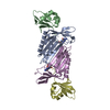
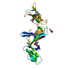
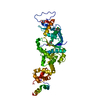
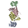
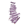
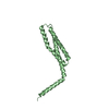
| タイトル | Crystal Structure of the N-terminal region of AlphaII-spectrin Tetramerization Domain |
|---|
 要素 要素 | Spectrin alpha chain, brain |
|---|
 キーワード キーワード | ACTIN BINDING / STRUCTURAL PROTEIN / lone helix followed by a triple helical bundle / Actin capping / Actin-binding / Alternative splicing / Calcium / Calmodulin-binding / Cytoplasm / Cytoskeleton / Phosphoprotein / Polymorphism / SH3 domain / SPECTRIN |
|---|
| 機能・相同性 |  機能・相同性情報 機能・相同性情報
spectrin / actin filament capping / Nephrin family interactions / Sensory processing of sound by outer hair cells of the cochlea / Interaction between L1 and Ankyrins / Sensory processing of sound by inner hair cells of the cochlea / RHOV GTPase cycle / cortical actin cytoskeleton / RHOU GTPase cycle / Caspase-mediated cleavage of cytoskeletal proteins ...spectrin / actin filament capping / Nephrin family interactions / Sensory processing of sound by outer hair cells of the cochlea / Interaction between L1 and Ankyrins / Sensory processing of sound by inner hair cells of the cochlea / RHOV GTPase cycle / cortical actin cytoskeleton / RHOU GTPase cycle / Caspase-mediated cleavage of cytoskeletal proteins / COPI-mediated anterograde transport / NCAM signaling for neurite out-growth / cell projection / structural constituent of cytoskeleton / specific granule lumen / actin filament binding / cell junction / tertiary granule lumen / extracellular vesicle / microtubule cytoskeleton / actin binding / RAF/MAP kinase cascade / actin cytoskeleton organization / calmodulin binding / cadherin binding / intracellular membrane-bounded organelle / calcium ion binding / Neutrophil degranulation / extracellular exosome / extracellular region / membrane / plasma membrane / cytosol類似検索 - 分子機能 Methane Monooxygenase Hydroxylase; Chain G, domain 1 - #60 / Alpha Spectrin, SH3 domain / EF-hand, Ca insensitive / Ca2+ insensitive EF hand / Ca2+ insensitive EF hand / Spectrin repeat / Spectrin repeat / Spectrin/alpha-actinin / Spectrin repeats / SH3 domain ...Methane Monooxygenase Hydroxylase; Chain G, domain 1 - #60 / Alpha Spectrin, SH3 domain / EF-hand, Ca insensitive / Ca2+ insensitive EF hand / Ca2+ insensitive EF hand / Spectrin repeat / Spectrin repeat / Spectrin/alpha-actinin / Spectrin repeats / SH3 domain / Methane Monooxygenase Hydroxylase; Chain G, domain 1 / EF-hand domain pair / EF-hand, calcium binding motif / Src homology 3 domains / SH3-like domain superfamily / EF-Hand 1, calcium-binding site / Src homology 3 (SH3) domain profile. / SH3 domain / EF-hand calcium-binding domain. / EF-hand calcium-binding domain profile. / EF-hand domain / EF-hand domain pair / Up-down Bundle / Mainly Alpha類似検索 - ドメイン・相同性 |
|---|
| 生物種 |  Homo sapiens (ヒト) Homo sapiens (ヒト) |
|---|
| 手法 |  X線回折 / 単波長異常分散 / 解像度: 2.3 Å X線回折 / 単波長異常分散 / 解像度: 2.3 Å |
|---|
 データ登録者 データ登録者 | Mehboob, S. / Santarsiero, B.D. / Long, F. / Witek, M. / Fung, L.W. |
|---|
 引用 引用 | ジャーナル: J.Biol.Chem. / 年: 2010
タイトル: Crystal structure of the nonerythroid alpha-spectrin tetramerization site reveals differences between erythroid and nonerythroid spectrin tetramer formation.
著者: Mehboob, S. / Song, Y. / Witek, M. / Long, F. / Santarsiero, B.D. / Johnson, M.E. / Fung, L.W. |
|---|
| 履歴 | | 登録 | 2008年10月30日 | 登録サイト: RCSB / 処理サイト: RCSB |
|---|
| 改定 1.0 | 2009年10月13日 | Provider: repository / タイプ: Initial release |
|---|
| 改定 1.1 | 2011年7月13日 | Group: Version format compliance |
|---|
| 改定 1.2 | 2017年10月25日 | Group: Author supporting evidence / カテゴリ: pdbx_struct_assembly_auth_evidence |
|---|
| 改定 1.3 | 2023年12月27日 | Group: Data collection / Database references
カテゴリ: chem_comp_atom / chem_comp_bond ...chem_comp_atom / chem_comp_bond / database_2 / struct_ref_seq_dif
Item: _database_2.pdbx_DOI / _database_2.pdbx_database_accession / _struct_ref_seq_dif.details |
|---|
|
|---|
 ムービー
ムービー コントローラー


 データを開く
 基本情報
基本情報 構造の表示
構造の表示 ダウンロードとリンク
 その他のダウンロード


 PDBj
PDBj


 集合体
集合体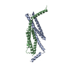


 分子量: 18.015 Da / 分子数: 206 / 由来タイプ: 天然 / 式: H2O
 試料調製
試料調製 解析
解析