 Movie
Movie Controller

[English] 日本語
 Yorodumi
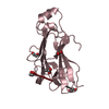
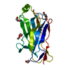
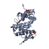
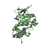
- PDB-3f2l: Crystal structure analysis of the K171A mutation of N-terminal ty... -
+ Open data

- Basic information
| Entry | Database: PDB / ID: 3f2l | ||||||
|---|---|---|---|---|---|---|---|
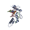
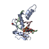
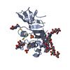
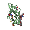
| Title | Crystal structure analysis of the K171A mutation of N-terminal type II cohesin 1 from the cellulosomal ScaB subunit of Acetivibrio cellulolyticus | ||||||
 Components Components | Cellulosomal scaffoldin adaptor protein B | ||||||
 Keywords Keywords | STRUCTURAL PROTEIN / Point mutation | ||||||
| Function / homology |  Function and homology information Function and homology informationpolysaccharide catabolic process / hydrolase activity, hydrolyzing O-glycosyl compounds / carbohydrate binding / extracellular region / metal ion binding Similarity search - Function | ||||||
| Biological species |  Acetivibrio cellulolyticus (bacteria) Acetivibrio cellulolyticus (bacteria) | ||||||
| Method |  X-RAY DIFFRACTION / FOURIER SYNTHESIS / Resolution: 1.85 Å X-RAY DIFFRACTION / FOURIER SYNTHESIS / Resolution: 1.85 Å | ||||||
 Authors Authors | Frolow, F. / Freeman, A. / Wine, Y. / Eppel, A. / Shanzer, M. / Stempler, S. | ||||||
 Citation Citation | Journal: To be Published Title: Crystal structure analysis of the K171A mutation of N-terminal type II cohesin 1 from the cellulosomal ScaB subunit of Acetivibrio cellulolyticus Authors: Frolow, F. / Freeman, A. / Wine, Y. / Eppel, A. / Shanzer, M. / Stempler, S. | ||||||
| History |
|
- Structure visualization
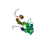
| Structure viewer | Molecule: MolmilJmol/JSmol |
|---|
- Downloads & links
-Download
| PDBx/mmCIF format | 3f2l.cif.gz | 93.5 KB | Display | PDBx/mmCIF format |
|---|---|---|---|---|
| PDB format | pdb3f2l.ent.gz | 70.9 KB | Display | PDB format |
| PDBx/mmJSON format | 3f2l.json.gz | Tree view | PDBx/mmJSON format | |
| Others |  Other downloads Other downloads |
-Validation report
| Arichive directory | https://data.pdbj.org/pub/pdb/validation_reports/f2/3f2lftp://data.pdbj.org/pub/pdb/validation_reports/f2/3f2l | HTTPS FTP |
|---|
-Related structure data
| Related structure data |  1qznS S: Starting model for refinement |
|---|---|
| Similar structure data |

-Links
 PDBj
PDBj

- Assembly
| Deposited unit | 
| ||||||||
|---|---|---|---|---|---|---|---|---|---|
| 1 |
| ||||||||
| Unit cell |
|
-Components
| #1: Protein | Mass: 18703.969 Da / Num. of mol.: 1 Fragment: N-terminal type II cohesin 1 from the cellulosomal ScaB subunit, UNP residues 28-199 Mutation: K171A Source method: isolated from a genetically manipulated source Source: (gene. exp.) Acetivibrio cellulolyticus (bacteria) / Plasmid: pET28a+ / Production host: | ||||||
|---|---|---|---|---|---|---|---|
| #2: Chemical | ChemComp-EDO /   Mass: 62.068 Da / Num. of mol.: 11 / Source method: obtained synthetically / Formula: C2H6O2 Mass: 62.068 Da / Num. of mol.: 11 / Source method: obtained synthetically / Formula: C2H6O2#3: Chemical | ChemComp-NH4 /   Mass: 18.038 Da / Num. of mol.: 8 / Source method: obtained synthetically / Formula: H4N Mass: 18.038 Da / Num. of mol.: 8 / Source method: obtained synthetically / Formula: H4N#4: Chemical | ChemComp-NO3 / |   Mass: 62.005 Da / Num. of mol.: 1 / Source method: obtained synthetically / Formula: NO3 Mass: 62.005 Da / Num. of mol.: 1 / Source method: obtained synthetically / Formula: NO3#5: Water | ChemComp-HOH / |  Mass: 18.015 Da / Num. of mol.: 239 / Source method: isolated from a natural source / Formula: H2O Mass: 18.015 Da / Num. of mol.: 239 / Source method: isolated from a natural source / Formula: H2O |
-Experimental details
-Experiment
| Experiment | Method: X-RAY DIFFRACTION / Number of used crystals: 1 |
|---|
- Sample preparation
| Crystal | Density Matthews: 2.52 Å3/Da / Density % sol: 51.21 % |
|---|---|
| Crystal grow | Temperature: 293 K / Method: microbatch / pH: 8.5 Details: 2.4M di-ammonium hydrogen phosphate, 0.1M Tris pH 8.5, Microbatch, temperature 293K |
-Data collection
| Diffraction | Mean temperature: 110 K |
|---|---|
| Diffraction source | Source: ROTATING ANODE / Type: RIGAKU MICROMAX-007 / Wavelength: 1.5418 Å |
| Detector | Type: RIGAKU RAXIS IV++ / Detector: IMAGE PLATE / Date: May 1, 2006 / Details: Osmic Confocal Mirrors |
| Radiation | Monochromator: Osmic Mirrors / Protocol: SINGLE WAVELENGTH / Monochromatic (M) / Laue (L): M / Scattering type: x-ray |
| Radiation wavelength | Wavelength: 1.5418 Å / Relative weight: 1 |
| Reflection | Resolution: 1.85→50 Å / Num. all: 16830 / Num. obs: 16830 / % possible obs: 100 % / Observed criterion σ(I): -3 / Redundancy: 5.7 % / Biso Wilson estimate: 27.53 Å2 / Rmerge(I) obs: 0.097 / Rsym value: 0.097 / Net I/σ(I): 11.4 |
| Reflection shell | Resolution: 1.85→1.85 Å / Redundancy: 5.7 % / Rmerge(I) obs: 0.49 / Mean I/σ(I) obs: 1.7 / Num. unique all: 16830 / Rsym value: 0.49 / % possible all: 100 |
- Processing
| Software |
| |||||||||||||||||||||||||||||||||||||||||||||||||||||||||||||||||||||||||||||||||||||||||||||||||||||||||
|---|---|---|---|---|---|---|---|---|---|---|---|---|---|---|---|---|---|---|---|---|---|---|---|---|---|---|---|---|---|---|---|---|---|---|---|---|---|---|---|---|---|---|---|---|---|---|---|---|---|---|---|---|---|---|---|---|---|---|---|---|---|---|---|---|---|---|---|---|---|---|---|---|---|---|---|---|---|---|---|---|---|---|---|---|---|---|---|---|---|---|---|---|---|---|---|---|---|---|---|---|---|---|---|---|---|---|
| Refinement | Method to determine structure: FOURIER SYNTHESIS Starting model: PDB ENTRY 1qzn Resolution: 1.85→29.21 Å / Cor.coef. Fo:Fc: 0.968 / Cor.coef. Fo:Fc free: 0.919 / SU B: 5.631 / SU ML: 0.078 / Isotropic thermal model: Anisotropic / Cross valid method: THROUGHOUT / ESU R: 0.313 / ESU R Free: 0.128 / Stereochemistry target values: MAXIMUM LIKELIHOOD / Details: HYDROGENS HAVE BEEN ADDED IN THE RIDING POSITIONS
| |||||||||||||||||||||||||||||||||||||||||||||||||||||||||||||||||||||||||||||||||||||||||||||||||||||||||
| Solvent computation | Ion probe radii: 0.8 Å / Shrinkage radii: 0.8 Å / VDW probe radii: 1.4 Å / Solvent model: BABINET MODEL WITH MASK | |||||||||||||||||||||||||||||||||||||||||||||||||||||||||||||||||||||||||||||||||||||||||||||||||||||||||
| Displacement parameters | Biso mean: 28.048 Å2
| |||||||||||||||||||||||||||||||||||||||||||||||||||||||||||||||||||||||||||||||||||||||||||||||||||||||||
| Refinement step | Cycle: LAST / Resolution: 1.85→29.21 Å
| |||||||||||||||||||||||||||||||||||||||||||||||||||||||||||||||||||||||||||||||||||||||||||||||||||||||||
| Refine LS restraints |
| |||||||||||||||||||||||||||||||||||||||||||||||||||||||||||||||||||||||||||||||||||||||||||||||||||||||||
| LS refinement shell | Resolution: 1.85→1.898 Å / Total num. of bins used: 20
|
