 Movie
Movie Controller

[English] 日本語
 Yorodumi
- PDB-3f0h: Crystal structure of Aminotransferase (RER070207000802) from Euba... -
+ Open data

- Basic information
| Entry | Database: PDB / ID: 3f0h | ||||||
|---|---|---|---|---|---|---|---|
| Title | Crystal structure of Aminotransferase (RER070207000802) from Eubacterium rectale at 1.70 A resolution | ||||||
 Components Components | Aminotransferase | ||||||
 Keywords Keywords | TRANSFERASE / RER070207000802 / Aminotransferase / Structural Genomics / Joint Center for Structural Genomics / JCSG / Protein Structure Initiative / PSI-2 / Aminotransferase class-V | ||||||
| Function / homology | Aspartate Aminotransferase, domain 1 / Aspartate Aminotransferase, domain 1 / Aspartate Aminotransferase; domain 2 / Type I PLP-dependent aspartate aminotransferase-like (Major domain) / Alpha-Beta Complex / 3-Layer(aba) Sandwich / Alpha Beta / Unknown ligand Function and homology information Function and homology information | ||||||
| Biological species |  Eubacterium rectale (bacteria) Eubacterium rectale (bacteria) | ||||||
| Method |  X-RAY DIFFRACTION / SYNCHROTRON / MAD / Resolution: 1.7 Å X-RAY DIFFRACTION / SYNCHROTRON / MAD / Resolution: 1.7 Å | ||||||
 Authors Authors | Joint Center for Structural Genomics (JCSG) | ||||||
 Citation Citation | Journal: To be published Title: Crystal structure of Aminotransferase (RER070207000802) from Eubacterium rectale at 1.70 A resolution Authors: Joint Center for Structural Genomics (JCSG) | ||||||
| History |
|
- Structure visualization
| Structure viewer | Molecule: MolmilJmol/JSmol |
|---|
- Downloads & links
-Download
| PDBx/mmCIF format | 3f0h.cif.gz | 100 KB | Display | PDBx/mmCIF format |
|---|---|---|---|---|
| PDB format | pdb3f0h.ent.gz | 74.4 KB | Display | PDB format |
| PDBx/mmJSON format | 3f0h.json.gz | Tree view | PDBx/mmJSON format | |
| Others |  Other downloads Other downloads |
-Validation report
| Arichive directory | https://data.pdbj.org/pub/pdb/validation_reports/f0/3f0hftp://data.pdbj.org/pub/pdb/validation_reports/f0/3f0h | HTTPS FTP |
|---|
-Related structure data
| Similar structure data | |
|---|---|
| Other databases |


-Links
 PDBj
PDBj

- Assembly
| Deposited unit | 
| ||||||||
|---|---|---|---|---|---|---|---|---|---|
| 1 | 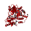
| ||||||||
| Unit cell |
|
-Components
| #1: Protein | Mass: 42970.754 Da / Num. of mol.: 1 Source method: isolated from a genetically manipulated source Source: (gene. exp.) Eubacterium rectale (bacteria) / Gene: RER070207000802 / Plasmid: SpeedET / Production host: | ||||
|---|---|---|---|---|---|
| #2: Chemical | ChemComp-UNL / Num. of mol.: 1 / Source method: obtained synthetically | ||||
| #3: Chemical |   Mass: 92.094 Da / Num. of mol.: 3 / Source method: obtained synthetically / Formula: C3H8O3 Mass: 92.094 Da / Num. of mol.: 3 / Source method: obtained synthetically / Formula: C3H8O3#4: Water | ChemComp-HOH / |  Mass: 18.015 Da / Num. of mol.: 377 / Source method: isolated from a natural source / Formula: H2O Mass: 18.015 Da / Num. of mol.: 377 / Source method: isolated from a natural source / Formula: H2OSequence details | THE CONSTRUCT WAS EXPRESSED WITH A PURIFICATI | |
-Experimental details
-Experiment
| Experiment | Method: X-RAY DIFFRACTION / Number of used crystals: 1 |
|---|
- Sample preparation
| Crystal | Density Matthews: 2.45 Å3/Da / Density % sol: 49.73 % |
|---|---|
| Crystal grow | Temperature: 277 K / Method: vapor diffusion, sitting drop / pH: 9 Details: 20.0000% PEG-6000, 0.1M Bicine pH 9.0, NANODROP, VAPOR DIFFUSION, SITTING DROP, temperature 277K |
-Data collection
| Diffraction | Mean temperature: 100 K | |||||||||||||||||||||||||||||||||||||||||||||||||||||||||||||||||||||||||||||
|---|---|---|---|---|---|---|---|---|---|---|---|---|---|---|---|---|---|---|---|---|---|---|---|---|---|---|---|---|---|---|---|---|---|---|---|---|---|---|---|---|---|---|---|---|---|---|---|---|---|---|---|---|---|---|---|---|---|---|---|---|---|---|---|---|---|---|---|---|---|---|---|---|---|---|---|---|---|---|
| Diffraction source | Source: SYNCHROTRON / Site: APS  / Beamline: 23-ID-B / Wavelength: 0.94645,0.97966 / Beamline: 23-ID-B / Wavelength: 0.94645,0.97966 | |||||||||||||||||||||||||||||||||||||||||||||||||||||||||||||||||||||||||||||
| Detector | Type: MARMOSAIC 300 mm CCD / Detector: CCD / Date: Oct 11, 2008 / Details: Adjustable focusing mirrors in K-B geometry | |||||||||||||||||||||||||||||||||||||||||||||||||||||||||||||||||||||||||||||
| Radiation | Monochromator: Si(111) Double Crystal Monochrometer / Protocol: MAD / Monochromatic (M) / Laue (L): M / Scattering type: x-ray | |||||||||||||||||||||||||||||||||||||||||||||||||||||||||||||||||||||||||||||
| Radiation wavelength |
| |||||||||||||||||||||||||||||||||||||||||||||||||||||||||||||||||||||||||||||
| Reflection | Resolution: 1.7→29.617 Å / Num. obs: 49360 / % possible obs: 99.6 % / Observed criterion σ(I): -3 / Redundancy: 20.28 % / Biso Wilson estimate: 23.322 Å2 / Rmerge(I) obs: 0.11 / Net I/σ(I): 13.56 | |||||||||||||||||||||||||||||||||||||||||||||||||||||||||||||||||||||||||||||
| Reflection shell |
|
-Phasing
| Phasing | Method: MAD |
|---|
- Processing
| Software |
| |||||||||||||||||||||||||||||||||||||||||||||||||||||||||||||||||||||||||||||||||||||
|---|---|---|---|---|---|---|---|---|---|---|---|---|---|---|---|---|---|---|---|---|---|---|---|---|---|---|---|---|---|---|---|---|---|---|---|---|---|---|---|---|---|---|---|---|---|---|---|---|---|---|---|---|---|---|---|---|---|---|---|---|---|---|---|---|---|---|---|---|---|---|---|---|---|---|---|---|---|---|---|---|---|---|---|---|---|---|
| Refinement | Method to determine structure: MAD / Resolution: 1.7→29.617 Å / Cor.coef. Fo:Fc: 0.973 / Cor.coef. Fo:Fc free: 0.969 / Occupancy max: 1 / Occupancy min: 0.3 / SU B: 3.394 / SU ML: 0.049 / TLS residual ADP flag: LIKELY RESIDUAL / Cross valid method: THROUGHOUT / σ(F): 0 / ESU R: 0.081 / ESU R Free: 0.078 Stereochemistry target values: MAXIMUM LIKELIHOOD WITH PHASES Details: 1. HYDROGENS HAVE BEEN ADDED IN THE RIDING POSITIONS. 2. A MET-INHIBITION PROTOCOL WAS USED FOR SELENOMETHIONINE INCORPORATION DURING PROTEIN EXPRESSION. THE OCCUPANCY OF THE SE ATOMS IN THE ...Details: 1. HYDROGENS HAVE BEEN ADDED IN THE RIDING POSITIONS. 2. A MET-INHIBITION PROTOCOL WAS USED FOR SELENOMETHIONINE INCORPORATION DURING PROTEIN EXPRESSION. THE OCCUPANCY OF THE SE ATOMS IN THE MSE RESIDUES WAS REDUCED TO 0.75 FOR THE REDUCED SCATTERING POWER DUE TO PARTIAL S-MET INCORPORATION. 3. ATOM RECORDS CONTAIN RESIDUAL B FACTORS ONLY. 4. PLP IS COVALENTLY ATTACHED TO LYS187 (LLP). RESIDUAL DENSITY SUGGESTS ADDITIONAL MOIETY MAY BE ATTACHED TO THE C4A ATOM OF PLP. THESE DENSITIES ARE MODELED AS AN UNKNOWN LIGAND (UNL). 5. GLYCEROL MODELED ARE PRESENT IN CRYO CONDITION.
| |||||||||||||||||||||||||||||||||||||||||||||||||||||||||||||||||||||||||||||||||||||
| Solvent computation | Ion probe radii: 0.8 Å / Shrinkage radii: 0.8 Å / VDW probe radii: 1.2 Å / Solvent model: MASK | |||||||||||||||||||||||||||||||||||||||||||||||||||||||||||||||||||||||||||||||||||||
| Displacement parameters | Biso max: 64.51 Å2 / Biso mean: 22.138 Å2 / Biso min: 8.97 Å2
| |||||||||||||||||||||||||||||||||||||||||||||||||||||||||||||||||||||||||||||||||||||
| Refinement step | Cycle: LAST / Resolution: 1.7→29.617 Å
| |||||||||||||||||||||||||||||||||||||||||||||||||||||||||||||||||||||||||||||||||||||
| Refine LS restraints |
| |||||||||||||||||||||||||||||||||||||||||||||||||||||||||||||||||||||||||||||||||||||
| LS refinement shell | Resolution: 1.699→1.743 Å / Total num. of bins used: 20
| |||||||||||||||||||||||||||||||||||||||||||||||||||||||||||||||||||||||||||||||||||||
| Refinement TLS params. | Method: refined / Origin x: 2.3974 Å / Origin y: 42.6825 Å / Origin z: 15.6357 Å
|
