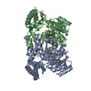
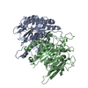
[English] 日本語
Yorodumi
- PDB-2ch2: Structure of the Anopheles gambiae 3-hydroxykynurenine transamina... -

+
Open data


ID or keywords:

Loading...

-
Basic information

Entry
Database: PDB / ID: 2ch2
TitleStructure of the Anopheles gambiae 3-hydroxykynurenine transaminase in complex with inhibitor
Components3-HYDROXYKYNURENINE TRANSAMINASE
KeywordsTRANSFERASE / PLP-ENZYME / KYNURENINE PATHWAY
Function / homology
Function and homology information


: / alanine-glyoxylate transaminase / L-kynurenine catabolic process / kynurenine-glyoxylate transaminase activity / kynurenine-glyoxylate transaminase / glycine biosynthetic process, by transamination of glyoxylate / serine-pyruvate transaminase activity / alanine-glyoxylate transaminase activity / glyoxylate catabolic process / pyridoxal phosphate binding / peroxisome
Similarity search - Function
Serine-pyruvate aminotransferase/2-aminoethylphosphonate-pyruvate transaminase / Aminotransferase class-V, pyridoxal-phosphate binding site / Aminotransferases class-V pyridoxal-phosphate attachment site. / Aminotransferase class V domain / Aminotransferase class-V / Aspartate Aminotransferase, domain 1 / Aspartate Aminotransferase, domain 1 / Aspartate Aminotransferase; domain 2 / Type I PLP-dependent aspartate aminotransferase-like (Major domain) / Pyridoxal phosphate-dependent transferase, small domain ...Serine-pyruvate aminotransferase/2-aminoethylphosphonate-pyruvate transaminase / Aminotransferase class-V, pyridoxal-phosphate binding site / Aminotransferases class-V pyridoxal-phosphate attachment site. / Aminotransferase class V domain / Aminotransferase class-V / Aspartate Aminotransferase, domain 1 / Aspartate Aminotransferase, domain 1 / Aspartate Aminotransferase; domain 2 / Type I PLP-dependent aspartate aminotransferase-like (Major domain) / Pyridoxal phosphate-dependent transferase, small domain / Pyridoxal phosphate-dependent transferase, major domain / Pyridoxal phosphate-dependent transferase / Alpha-Beta Complex / 3-Layer(aba) Sandwich / Alpha Beta
Similarity search - Domain/homology
4-(2-AMINOPHENYL)-4-OXOBUTANOIC ACID / PYRIDOXAL-5'-PHOSPHATE / 3-hydroxykynurenine transaminase / 3-hydroxykynurenine transaminase
Similarity search - Component
Biological speciesANOPHELES GAMBIAE (African malaria mosquito)
MethodX-RAY DIFFRACTION / SYNCHROTRON / OTHER / Resolution: 2.7 Å
AuthorsRossi, F. / Garavaglia, S. / Giovenzana, G.B. / Arca, B. / Li, J. / Rizzi, M.
CitationJournal: Proc.Natl.Acad.Sci.USA / Year: 2006
Title: Crystal Structure of the Anopheles Gambiae 3-Hydroxykynurenine Transaminase
Authors: Rossi, F. / Garavaglia, S. / Giovenzana, G.B. / Arca, B. / Li, J. / Rizzi, M.
History
DepositionMar 10, 2006Deposition site: PDBE / Processing site: PDBE
Revision 1.0Mar 28, 2006Provider: repository / Type: Initial release
Revision 1.1Jul 17, 2013Group: Atomic model / Database references ...Atomic model / Database references / Derived calculations / Non-polymer description / Other / Source and taxonomy / Structure summary / Version format compliance

-
Structure visualization

Structure viewerMolecule:
MolmilJmol/JSmol

Downloads & links

-
Assembly

Deposited unit
A: 3-HYDROXYKYNURENINE TRANSAMINASE
B: 3-HYDROXYKYNURENINE TRANSAMINASE
C: 3-HYDROXYKYNURENINE TRANSAMINASE
D: 3-HYDROXYKYNURENINE TRANSAMINASE
hetero molecules


Theoretical massNumber of molelcules
Total (without water)179,16212
Polymers177,4004
Non-polymers1,7618
Water2,270126
1
A: 3-HYDROXYKYNURENINE TRANSAMINASE
D: 3-HYDROXYKYNURENINE TRANSAMINASE
hetero molecules


Theoretical massNumber of molelcules
Total (without water)89,5816
Polymers88,7002
Non-polymers8814
Water362
TypeNameSymmetry operationNumber
identity operation1_555x,y,z1
Buried area8920 Å2
ΔGint-48.3 kcal/mol
Surface area26210 Å2
MethodPISA
2
B: 3-HYDROXYKYNURENINE TRANSAMINASE
C: 3-HYDROXYKYNURENINE TRANSAMINASE
hetero molecules


Theoretical massNumber of molelcules
Total (without water)89,5816
Polymers88,7002
Non-polymers8814
Water362
TypeNameSymmetry operationNumber
identity operation1_555x,y,z1
Buried area9010 Å2
ΔGint-46.3 kcal/mol
Surface area26140 Å2
MethodPISA
Unit cell
Length a, b, c (Å)87.940, 83.747, 118.655
Angle α, β, γ (deg.)90.00, 100.09, 90.00
Int Tables number4
Space group name H-MP1211
Noncrystallographic symmetry (NCS)NCS domain:
IDEns-IDDetails
11A
21B
31C
41D

NCS domain segments:
Dom-IDComponent-IDEns-IDRefine codeAuth asym-IDAuth seq-ID
1111A3 - 389
2111B3 - 389
3111C3 - 389
4111D3 - 389

-
Components

#1: Protein
3-HYDROXYKYNURENINE TRANSAMINASE


Mass: 44350.094 Da / Num. of mol.: 4
Source method: isolated from a genetically manipulated source
Source: (gene. exp.) ANOPHELES GAMBIAE (African malaria mosquito)
Strain: GASUA / Plasmid: PET16B / Production host: ESCHERICHIA COLI (E. coli) / Strain (production host): BL21(DE3) / References: UniProt: Q4LAM2, UniProt: Q7PRG3*PLUS
#2: Chemical
ChemComp-PLP / PYRIDOXAL-5'-PHOSPHATE / VITAMIN B6 Phosphate


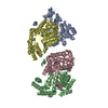
Mass: 247.142 Da / Num. of mol.: 4 / Source method: obtained synthetically / Formula: C8H10NO6P
#3: Chemical
ChemComp-KY1 / 4-(2-AMINOPHENYL)-4-OXOBUTANOIC ACID


Mass: 193.199 Da / Num. of mol.: 4 / Source method: obtained synthetically / Formula: C10H11NO3
#4: Water ChemComp-HOH / water


Mass: 18.015 Da / Num. of mol.: 126 / Source method: isolated from a natural source / Formula: H2O

-
Experimental details

-
Experiment

ExperimentMethod: X-RAY DIFFRACTION / Number of used crystals: 1

-
Sample preparation

CrystalDensity Matthews: 2.43 Å3/Da / Density % sol: 50 %
Crystal growpH: 7 / Details: pH 7.00

-
Data collection

DiffractionMean temperature: 100 K
Diffraction sourceSource: SYNCHROTRON / Site: ESRF / Beamline: ID14-1 / Wavelength: 1
DetectorType: ADSC CCD / Detector: CCD / Date: Feb 13, 2005
RadiationProtocol: SINGLE WAVELENGTH / Monochromatic (M) / Laue (L): M / Scattering type: x-ray
Radiation wavelengthWavelength: 1 Å / Relative weight: 1
ReflectionResolution: 2.7→20 Å / Num. obs: 45886 / % possible obs: 98.2 % / Observed criterion σ(I): 0 / Redundancy: 2.7 % / Rmerge(I) obs: 0.07 / Net I/σ(I): 12.5
Reflection shellResolution: 2.5→2.7 Å / Redundancy: 2.5 % / Rmerge(I) obs: 0.11 / Mean I/σ(I) obs: 3 / % possible all: 97.4

-
Processing

SoftwareName: REFMAC / Version: 5.2.0005 / Classification: refinement
RefinementMethod to determine structure: OTHER / Resolution: 2.7→50 Å / Cor.coef. Fo:Fc: 0.923 / Cor.coef. Fo:Fc free: 0.887 / SU B: 14.752 / SU ML: 0.292 / Cross valid method: THROUGHOUT / ESU R Free: 0.379 / Stereochemistry target values: MAXIMUM LIKELIHOOD
Details: HYDROGENS HAVE BEEN ADDED IN THE RIDING POSITIONS. RESIDUES 1-2 AND 390-396 WERE NOT VISIBLE IN THE ELECTRON DENSITY AND NOT INCLUDED IN THE FINAL MODEL.
RfactorNum. reflection% reflectionSelection details
Rfree0.266 979 2.1 %RANDOM
Rwork0.213 ---
obs0.214 44886 100 %-
Solvent computationIon probe radii: 0.8 Å / Shrinkage radii: 0.8 Å / VDW probe radii: 1.2 Å / Solvent model: BABINET MODEL WITH MASK
Displacement parametersBiso mean: 37.52 Å2
Baniso -1Baniso -2Baniso -3
1--0.4 Å20 Å2-1.13 Å2
2--3.41 Å20 Å2
3----3.41 Å2
Refinement stepCycle: LAST / Resolution: 2.7→50 Å
ProteinNucleic acidLigandSolventTotal
Num. atoms12143 0 116 126 12385
Refine LS restraints
Refine-IDTypeDev idealDev ideal targetNumber
X-RAY DIFFRACTIONr_bond_refined_d0.0170.02212548
X-RAY DIFFRACTIONr_bond_other_d
X-RAY DIFFRACTIONr_angle_refined_deg1.6021.97617017
X-RAY DIFFRACTIONr_angle_other_deg
X-RAY DIFFRACTIONr_dihedral_angle_1_deg6.17151544
X-RAY DIFFRACTIONr_dihedral_angle_2_deg38.11823.502534
X-RAY DIFFRACTIONr_dihedral_angle_3_deg17.953152123
X-RAY DIFFRACTIONr_dihedral_angle_4_deg21.8651584
X-RAY DIFFRACTIONr_chiral_restr0.1070.21848
X-RAY DIFFRACTIONr_gen_planes_refined0.0050.029494
X-RAY DIFFRACTIONr_gen_planes_other
X-RAY DIFFRACTIONr_nbd_refined0.2310.25994
X-RAY DIFFRACTIONr_nbd_other
X-RAY DIFFRACTIONr_nbtor_refined0.3220.28703
X-RAY DIFFRACTIONr_nbtor_other
X-RAY DIFFRACTIONr_xyhbond_nbd_refined0.1480.2460
X-RAY DIFFRACTIONr_xyhbond_nbd_other
X-RAY DIFFRACTIONr_metal_ion_refined
X-RAY DIFFRACTIONr_metal_ion_other
X-RAY DIFFRACTIONr_symmetry_vdw_refined0.3550.2108
X-RAY DIFFRACTIONr_symmetry_vdw_other
X-RAY DIFFRACTIONr_symmetry_hbond_refined0.5610.23
X-RAY DIFFRACTIONr_symmetry_hbond_other
X-RAY DIFFRACTIONr_symmetry_metal_ion_refined
X-RAY DIFFRACTIONr_symmetry_metal_ion_other
X-RAY DIFFRACTIONr_mcbond_it0.7511.57846
X-RAY DIFFRACTIONr_mcbond_other
X-RAY DIFFRACTIONr_mcangle_it1.202212430
X-RAY DIFFRACTIONr_mcangle_other
X-RAY DIFFRACTIONr_scbond_it1.88735348
X-RAY DIFFRACTIONr_scbond_other
X-RAY DIFFRACTIONr_scangle_it3.2344.54587
X-RAY DIFFRACTIONr_scangle_other
X-RAY DIFFRACTIONr_long_range_B_refined
X-RAY DIFFRACTIONr_long_range_B_other
X-RAY DIFFRACTIONr_rigid_bond_restr
X-RAY DIFFRACTIONr_sphericity_free
X-RAY DIFFRACTIONr_sphericity_bonded
Refine LS restraints NCS

Ens-ID: 1 / Number: 3033 / Refine-ID: X-RAY DIFFRACTION

Dom-IDAuth asym-IDTypeRms dev position (Å)Weight position
1Atight positional0.080.05
2Btight positional0.070.05
3Ctight positional0.080.05
4Dtight positional0.090.05
1Atight thermal0.170.5
2Btight thermal0.180.5
3Ctight thermal0.160.5
4Dtight thermal0.220.5
LS refinement shellResolution: 2.7→2.77 Å / Total num. of bins used: 20 /
RfactorNum. reflection
Rfree0.483 63
Rwork0.336 3345

+
About Yorodumi

-
News

-
Feb 9, 2022. New format data for meta-information of EMDB entries

New format data for meta-information of EMDB entries

  • Version 3 of the EMDB header file is now the official format.
  • The previous official version 1.9 will be removed from the archive.

Related info.:EMDB header

External links:wwPDB to switch to version 3 of the EMDB data model

-
Aug 12, 2020. Covid-19 info

Covid-19 info

URL: https://pdbjlc1.pdbj.org/emnavi/covid19.php

New page: Covid-19 featured information page in EM Navigator.

Related info.:Covid-19 info / Mar 5, 2020. Novel coronavirus structure data

+
Mar 5, 2020. Novel coronavirus structure data

Novel coronavirus structure data

Related info.:Yorodumi Speices / Aug 12, 2020. Covid-19 info

External links:COVID-19 featured content - PDBj / Molecule of the Month (242):Coronavirus Proteases

+
Jan 31, 2019. EMDB accession codes are about to change! (news from PDBe EMDB page)

EMDB accession codes are about to change! (news from PDBe EMDB page)

  • The allocation of 4 digits for EMDB accession codes will soon come to an end. Whilst these codes will remain in use, new EMDB accession codes will include an additional digit and will expand incrementally as the available range of codes is exhausted. The current 4-digit format prefixed with “EMD-” (i.e. EMD-XXXX) will advance to a 5-digit format (i.e. EMD-XXXXX), and so on. It is currently estimated that the 4-digit codes will be depleted around Spring 2019, at which point the 5-digit format will come into force.
  • The EM Navigator/Yorodumi systems omit the EMD- prefix.

Related info.:Q: What is EMD? / ID/Accession-code notation in Yorodumi/EM Navigator

External links:EMDB Accession Codes are Changing Soon! / Contact to PDBj

+
Jul 12, 2017. Major update of PDB

Major update of PDB

  • wwPDB released updated PDB data conforming to the new PDBx/mmCIF dictionary.
  • This is a major update changing the version number from 4 to 5, and with Remediation, in which all the entries are updated.
  • In this update, many items about electron microscopy experimental information are reorganized (e.g. em_software).
  • Now, EM Navigator and Yorodumi are based on the updated data.

External links:wwPDB Remediation / Enriched Model Files Conforming to OneDep Data Standards Now Available in the PDB FTP Archive

-
Yorodumi

Thousand views of thousand structures

  • Yorodumi is a browser for structure data from EMDB, PDB, SASBDB, etc.
  • This page is also the successor to EM Navigator detail page, and also detail information page/front-end page for Omokage search.
  • The word "yorodu" (or yorozu) is an old Japanese word meaning "ten thousand". "mi" (miru) is to see.

Related info.:EMDB / PDB / SASBDB / Comparison of 3 databanks / Yorodumi Search / Aug 31, 2016. New EM Navigator & Yorodumi / Yorodumi Papers / Jmol/JSmol / Function and homology information / Changes in new EM Navigator and Yorodumi

Read more