Mass: 18.015 Da / Num. of mol.: 564 / Source method: isolated from a natural source / Formula: H2O
Sequence details
THE CONSTRUCT WAS EXPRESSED WITH A PURIFICATION TAG MGSDKIHHHHHHENLYFQG. THE TAG WAS REMOVED WITH ...THE CONSTRUCT WAS EXPRESSED WITH A PURIFICATION TAG MGSDKIHHHHHHENLYFQG. THE TAG WAS REMOVED WITH TEV PROTEASE LEAVING ONLY A GLYCINE (0) FOLLOWED BY THE TARGET SEQUENCE.
-
Experimental details
-
Experiment
Experiment
Method: X-RAY DIFFRACTION / Number of used crystals: 1
-
Sample preparation
Crystal
Density Matthews: 2.12 Å3/Da / Density % sol: 42.07 %
Crystal grow
Temperature: 277 K / Method: vapor diffusion, sitting drop / pH: 7.5 Details: NANODROP, 10.0% Isopropanol, 20.0% PEG 4000, 0.1M HEPES pH 7.5, VAPOR DIFFUSION, SITTING DROP, temperature 277K
-
Data collection
Diffraction
Mean temperature: 100 K
Diffraction source
Source: SYNCHROTRON / Site: ALS / Beamline: 8.2.2 / Wavelength: 0.9789 Å
Monochromator: Double Crystal Si(111) / Protocol: SAD / Monochromatic (M) / Laue (L): M / Scattering type: x-ray
Radiation wavelength
Wavelength: 0.9789 Å / Relative weight: 1
Reflection
Resolution: 2.01→45.549 Å / Num. obs: 46287 / % possible obs: 95.3 % / Observed criterion σ(I): -3 / Biso Wilson estimate: 23.545 Å2 / Rmerge(I) obs: 0.085
Reflection shell
Resolution (Å)
Rmerge(I) obs
Mean I/σ(I) obs
Num. measured obs
Num. unique obs
Diffraction-ID
% possible all
2.01-2.08
0.525
2
10011
3549
1
75.9
2.08-2.17
0.411
2.6
14015
4743
1
92.3
2.17-2.26
0.31
3.6
12247
4056
1
93
2.26-2.38
0.278
3.9
14832
4628
1
95.5
2.38-2.53
0.21
5.4
16021
4764
1
98.1
2.53-2.73
0.157
7.1
17098
4909
1
99.4
2.73-3
0.109
9.5
16929
4761
1
99.8
3-3.43
0.068
13.6
17226
4836
1
99.8
3.43-4.32
0.044
17.9
16972
4888
1
99.1
4.32-45.549
0.04
19.6
17282
5153
1
99
-
Phasing
Phasing
Method: SAD
-
Processing
Software
Name
Version
Classification
NB
REFMAC
5.2.0019
refinement
PHENIX
refinement
SHELX
phasing
MolProbity
3beta29
modelbuilding
XSCALE
datascaling
PDB_EXTRACT
3.006
dataextraction
ADSC
Quantum
datacollection
XDS
datareduction
SHELXD
phasing
autoSHARP
phasing
Refinement
Method to determine structure: SAD / Resolution: 2.01→45.549 Å / Cor.coef. Fo:Fc: 0.959 / Cor.coef. Fo:Fc free: 0.923 / Occupancy max: 1 / Occupancy min: 0.25 / SU B: 10.067 / SU ML: 0.141 / TLS residual ADP flag: LIKELY RESIDUAL / Cross valid method: THROUGHOUT / σ(F): 0 / ESU R: 0.211 / ESU R Free: 0.185 Stereochemistry target values: MAXIMUM LIKELIHOOD WITH PHASES Details: 1. HYDROGENS HAVE BEEN ADDED IN THE RIDING POSITIONS. 2. ATOM RECORD CONTAINS RESIDUAL B FACTORS ONLY. 3. A MET-INHIBITION PROTOCOL WAS USED FOR SELENOMETHIONINE INCORPORATION DURING PROTEIN ...Details: 1. HYDROGENS HAVE BEEN ADDED IN THE RIDING POSITIONS. 2. ATOM RECORD CONTAINS RESIDUAL B FACTORS ONLY. 3. A MET-INHIBITION PROTOCOL WAS USED FOR SELENOMETHIONINE INCORPORATION DURING PROTEIN EXPRESSION. THE OCCUPANCY OF THE SE ATOMS IN THE MSE RESIDUES WAS REDUCED TO 0.75 TO ACCOUNT FOR THE REDUCED SCATTERING POWER DUE TO PARTIAL S-MET INCORPORATION. 4. RESIDUE LYSINE 226 IN BOTH CHAINS HAS BEEN MODELED AS LLP - A LYSINE WITH A SCHIFF BASE LINKAGE TO PYRIDOXAL-5'PHOSPHATE (PLP). THIS IS THE CONSERVED ACTIVE SITE. 5. 2-PROPANOL AND GLYCEROL MOLECULES FROM CRYSTALLIZATION AND CRYOPROTECTANT WERE MODELED IN THE STRUCTURE, RESPECTIVELY.
Rfactor
Num. reflection
% reflection
Selection details
Rfree
0.235
2335
5.1 %
RANDOM
Rwork
0.174
-
-
-
obs
0.177
46226
95.35 %
-
Solvent computation
Ion probe radii: 0.8 Å / Shrinkage radii: 0.8 Å / VDW probe radii: 1.2 Å / Solvent model: BABINET MODEL WITH MASK
In the structure databanks used in Yorodumi, some data are registered as the other names, "COVID-19 virus" and "2019-nCoV". Here are the details of the virus and the list of structure data.
Jan 31, 2019. EMDB accession codes are about to change! (news from PDBe EMDB page)
EMDB accession codes are about to change! (news from PDBe EMDB page)
The allocation of 4 digits for EMDB accession codes will soon come to an end. Whilst these codes will remain in use, new EMDB accession codes will include an additional digit and will expand incrementally as the available range of codes is exhausted. The current 4-digit format prefixed with “EMD-” (i.e. EMD-XXXX) will advance to a 5-digit format (i.e. EMD-XXXXX), and so on. It is currently estimated that the 4-digit codes will be depleted around Spring 2019, at which point the 5-digit format will come into force.
The EM Navigator/Yorodumi systems omit the EMD- prefix.
Related info.:Q: What is EMD? / ID/Accession-code notation in Yorodumi/EM Navigator
Yorodumi is a browser for structure data from EMDB, PDB, SASBDB, etc.
This page is also the successor to EM Navigator detail page, and also detail information page/front-end page for Omokage search.
The word "yorodu" (or yorozu) is an old Japanese word meaning "ten thousand". "mi" (miru) is to see.
Related info.:EMDB / PDB / SASBDB / Comparison of 3 databanks / Yorodumi Search / Aug 31, 2016. New EM Navigator & Yorodumi / Yorodumi Papers / Jmol/JSmol / Function and homology information / Changes in new EM Navigator and Yorodumi
 Movie
Movie Controller

 Yorodumi
 Open data
 Basic information
 Components
 Keywords
 Function and homology information
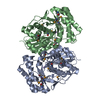
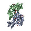
 Campylobacter jejuni subsp. jejuni NCTC 11168 (Campylobacter)
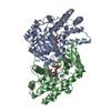
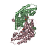
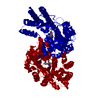
 X-RAY DIFFRACTION /
  Authors
 Citation
 Structure visualization
 Downloads & links
 Other downloads
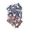


 PDBj
PDBj
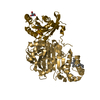
 Assembly

 Mass: 60.095 Da / Num. of mol.: 2 / Source method: obtained synthetically / Formula: C3H8O
 Mass: 92.094 Da / Num. of mol.: 1 / Source method: obtained synthetically / Formula: C3H8O3
 Mass: 18.015 Da / Num. of mol.: 564 / Source method: isolated from a natural source / Formula: H2O
 Sample preparation
 / Beamline: 8.2.2 / Wavelength: 0.9789 Å
 Processing