 Movie
Movie Controller

[English] 日本語
 Yorodumi
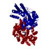
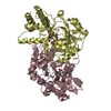
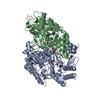
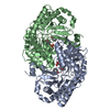
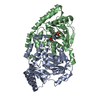
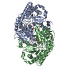
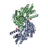
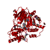
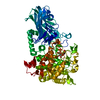
- PDB-3dxv: The crystal structure of alpha-amino-epsilon-caprolactam racemase... -
+ Open data

- Basic information
| Entry | Database: PDB / ID: 3dxv | ||||||
|---|---|---|---|---|---|---|---|
| Title | The crystal structure of alpha-amino-epsilon-caprolactam racemase from Achromobacter obae | ||||||
 Components Components | Alpha-amino-epsilon-caprolactam racemase | ||||||
 Keywords Keywords | ISOMERASE / fold-type1 / pyridoxal-5'-phosphate dependent racemase / Pyridoxal phosphate | ||||||
| Function / homology |  Function and homology information Function and homology information2-aminohexano-6-lactam racemase / 2-aminohexano-6-lactam racemase activity / pyridoxal phosphate binding Similarity search - Function | ||||||
| Biological species |  Achromobacter obae (bacteria) Achromobacter obae (bacteria) | ||||||
| Method |  X-RAY DIFFRACTION / SYNCHROTRON / MOLECULAR REPLACEMENT / Resolution: 2.21 Å X-RAY DIFFRACTION / SYNCHROTRON / MOLECULAR REPLACEMENT / Resolution: 2.21 Å | ||||||
 Authors Authors | Okazaki, S. / Suzuki, A. / Komeda, H. / Asano, Y. / Yamane, T. | ||||||
 Citation Citation | Journal: Biochemistry / Year: 2009 Title: The novel structure of a pyridoxal 5'-phosphate-dependent fold-type I racemase, alpha-amino-epsilon-caprolactam racemase from Achromobacter obae Authors: Okazaki, S. / Suzuki, A. / Mizushima, T. / Kawano, T. / Komeda, H. / Asano, Y. / Yamane, T. | ||||||
| History |
|
- Structure visualization
| Structure viewer | Molecule: MolmilJmol/JSmol |
|---|
- Downloads & links
-Download
| PDBx/mmCIF format | 3dxv.cif.gz | 168.4 KB | Display | PDBx/mmCIF format |
|---|---|---|---|---|
| PDB format | pdb3dxv.ent.gz | 132.8 KB | Display | PDB format |
| PDBx/mmJSON format | 3dxv.json.gz | Tree view | PDBx/mmJSON format | |
| Others |  Other downloads Other downloads |
-Validation report
| Arichive directory | https://data.pdbj.org/pub/pdb/validation_reports/dx/3dxvftp://data.pdbj.org/pub/pdb/validation_reports/dx/3dxv | HTTPS FTP |
|---|
-Related structure data
| Related structure data |  2zukC  3dxwC  1sftS C: citing same article ( S: Starting model for refinement |
|---|---|
| Similar structure data |

-Links
 PDBj
PDBj- Assembly
| Deposited unit | 
| ||||||||
|---|---|---|---|---|---|---|---|---|---|
| 1 |
| ||||||||
| Unit cell |
|
-Components
| #1: Protein | Mass: 46031.473 Da / Num. of mol.: 2 Source method: isolated from a genetically manipulated source Source: (gene. exp.) Achromobacter obae (bacteria) / Plasmid: pET15b / Production host: References: UniProt: Q7M181, 2-aminohexano-6-lactam racemase #2: Chemical |   Mass: 247.142 Da / Num. of mol.: 2 / Source method: obtained synthetically / Formula: C8H10NO6P Mass: 247.142 Da / Num. of mol.: 2 / Source method: obtained synthetically / Formula: C8H10NO6P#3: Water | ChemComp-HOH / |  Mass: 18.015 Da / Num. of mol.: 120 / Source method: isolated from a natural source / Formula: H2O Mass: 18.015 Da / Num. of mol.: 120 / Source method: isolated from a natural source / Formula: H2O |
|---|
-Experimental details
-Experiment
| Experiment | Method: X-RAY DIFFRACTION / Number of used crystals: 1 |
|---|
- Sample preparation
| Crystal | Density Matthews: 1.86 Å3/Da / Density % sol: 33.85 % |
|---|---|
| Crystal grow | Temperature: 293 K / Method: vapor diffusion, hanging drop / pH: 8.7 Details: 25% PEG4000, 0.17M magnesium chloride, 0.08M Tris/HCl (pH8.7), 5% Sucrose, VAPOR DIFFUSION, HANGING DROP, temperature 293K |
-Data collection
| Diffraction | Mean temperature: 100 K |
|---|---|
| Diffraction source | Source: SYNCHROTRON / Site: NSRRC  / Beamline: BL13B1 / Wavelength: 1 Å / Beamline: BL13B1 / Wavelength: 1 Å |
| Detector | Type: ADSC QUANTUM 315 / Detector: CCD / Date: Mar 14, 2008 |
| Radiation | Monochromator: LN2-cooled, fixed-exit double crystal monochromator Protocol: SINGLE WAVELENGTH / Monochromatic (M) / Laue (L): M / Scattering type: x-ray |
| Radiation wavelength | Wavelength: 1 Å / Relative weight: 1 |
| Reflection | Resolution: 2.21→50 Å / Num. obs: 35281 / % possible obs: 99.6 % / Observed criterion σ(I): 0 / Redundancy: 5.9 % / Rmerge(I) obs: 0.108 / Rsym value: 0.074 |
| Reflection shell | Resolution: 2.21→2.29 Å / Redundancy: 5.2 % / Rmerge(I) obs: 0.494 / Rsym value: 0.429 / % possible all: 98.2 |
- Processing
| Software |
| ||||||||||||||||||||||||||||||||||||||||||||||||||||||||||||||||||||||||||||||||||||||||||
|---|---|---|---|---|---|---|---|---|---|---|---|---|---|---|---|---|---|---|---|---|---|---|---|---|---|---|---|---|---|---|---|---|---|---|---|---|---|---|---|---|---|---|---|---|---|---|---|---|---|---|---|---|---|---|---|---|---|---|---|---|---|---|---|---|---|---|---|---|---|---|---|---|---|---|---|---|---|---|---|---|---|---|---|---|---|---|---|---|---|---|---|
| Refinement | Method to determine structure: MOLECULAR REPLACEMENT Starting model: PDB ENTRY 1SFT Resolution: 2.21→19.96 Å / Cor.coef. Fo:Fc: 0.949 / Cor.coef. Fo:Fc free: 0.922 / SU B: 7.74 / SU ML: 0.192 / Cross valid method: THROUGHOUT / σ(F): 0 / ESU R: 0.412 / ESU R Free: 0.243 / Stereochemistry target values: MAXIMUM LIKELIHOOD / Details: HYDROGENS HAVE BEEN ADDED IN THE RIDING POSITIONS
| ||||||||||||||||||||||||||||||||||||||||||||||||||||||||||||||||||||||||||||||||||||||||||
| Solvent computation | Ion probe radii: 0.8 Å / Shrinkage radii: 0.8 Å / VDW probe radii: 1.2 Å / Solvent model: MASK | ||||||||||||||||||||||||||||||||||||||||||||||||||||||||||||||||||||||||||||||||||||||||||
| Displacement parameters | Biso mean: 30.541 Å2
| ||||||||||||||||||||||||||||||||||||||||||||||||||||||||||||||||||||||||||||||||||||||||||
| Refinement step | Cycle: LAST / Resolution: 2.21→19.96 Å
| ||||||||||||||||||||||||||||||||||||||||||||||||||||||||||||||||||||||||||||||||||||||||||
| Refine LS restraints |
| ||||||||||||||||||||||||||||||||||||||||||||||||||||||||||||||||||||||||||||||||||||||||||
| LS refinement shell | Resolution: 2.207→2.264 Å / Total num. of bins used: 20
|
