- PDB-3ezs: Crystal structure of aminotransferase AspB (NP_207418.1) from HEL... -
+
Open data
ID or keywords:
Loading...
-
Basic information
Entry
Database: PDB / ID: 3ezs
Title
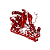
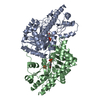
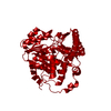
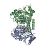
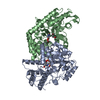
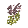
Crystal structure of aminotransferase AspB (NP_207418.1) from HELICOBACTER PYLORI 26695 at 2.19 A resolution
Components
aminotransferase AspB
Keywords
TRANSFERASE / NP_207418.1 / aminotransferase AspB / Structural Genomics / Joint Center for Structural Genomics / JCSG / Protein Structure Initiative / PSI-2 / Aminotransferase class I and II
Mass: 18.015 Da / Num. of mol.: 402 / Source method: isolated from a natural source / Formula: H2O
Has protein modification
Y
Sequence details
SEQUENCE THE CONSTRUCT WAS EXPRESSED WITH A PURIFICATION TAG MGSDKIHHHHHHENLYFQG. THE TAG WAS ...SEQUENCE THE CONSTRUCT WAS EXPRESSED WITH A PURIFICATION TAG MGSDKIHHHHHHENLYFQG. THE TAG WAS REMOVED WITH TEV PROTEASE LEAVING ONLY A GLYCINE (0) FOLLOWED BY THE TARGET SEQUENCE.
-
Experimental details
-
Experiment
Experiment
Method: X-RAY DIFFRACTION / Number of used crystals: 1
-
Sample preparation
Crystal
Density Matthews: 2.28 Å3/Da / Density % sol: 45.94 %
Crystal grow
Temperature: 277 K / Method: vapor diffusion, sitting drop / pH: 7.5 Details: 15.0000% Glycerol, 8.5000% iso-Propanol, 17.0000% PEG-4000, 0.1M HEPES pH 7.5, NANODROP, VAPOR DIFFUSION, SITTING DROP, temperature 277K
Resolution: 2.19→44.281 Å / Num. obs: 40833 / % possible obs: 98.3 % / Observed criterion σ(I): -3 / Redundancy: 5.3 % / Biso Wilson estimate: 29.514 Å2 / Rmerge(I) obs: 0.112 / Net I/σ(I): 12.8
Reflection shell
Resolution (Å)
Rmerge(I) obs
Mean I/σ(I) obs
Num. measured obs
Num. unique obs
Diffraction-ID
% possible all
2.19-2.27
0.612
2.29
12871
3568
1
85.8
2.27-2.36
0.55
3.2
22277
4039
1
100
2.36-2.47
0.447
4
23107
4193
1
100
2.47-2.6
0.362
4.9
22471
4070
1
100
2.6-2.76
0.263
6.5
22276
4042
1
100
2.76-2.97
0.193
8.7
22492
4083
1
100
2.97-3.27
0.126
12.8
22750
4148
1
100
3.27-3.74
0.076
20.2
22609
4152
1
100
3.74-4.7
0.045
29.8
22659
4193
1
99.7
4.7-44.281
0.041
32.4
23010
4373
1
98.1
-
Phasing
Phasing
Method: MAD
-
Processing
Software
Name
Version
Classification
NB
REFMAC
5.2.0019
refinement
PHENIX
refinement
SHELX
phasing
MolProbity
3beta29
modelbuilding
XSCALE
datascaling
PDB_EXTRACT
3.006
dataextraction
XDS
datareduction
SHELXD
phasing
autoSHARP
phasing
Refinement
Method to determine structure: MAD / Resolution: 2.19→44.281 Å / Cor.coef. Fo:Fc: 0.963 / Cor.coef. Fo:Fc free: 0.93 / Occupancy max: 1 / Occupancy min: 0.37 / SU B: 10.513 / SU ML: 0.139 / TLS residual ADP flag: LIKELY RESIDUAL / Cross valid method: THROUGHOUT / σ(F): 0 / ESU R: 0.256 / ESU R Free: 0.198 Stereochemistry target values: MAXIMUM LIKELIHOOD WITH PHASES Details: 1. HYDROGENS HAVE BEEN ADDED IN RIDING POSITIONS. 2. ATOM RECORD CONTAINS RESIDUAL B FACTORS ONLY. 3. A MET-INHIBITION PROTOCOL WAS USED FOR SELENOMETHIONINE INCORPORATION DURING PROTEIN ...Details: 1. HYDROGENS HAVE BEEN ADDED IN RIDING POSITIONS. 2. ATOM RECORD CONTAINS RESIDUAL B FACTORS ONLY. 3. A MET-INHIBITION PROTOCOL WAS USED FOR SELENOMETHIONINE INCORPORATION DURING PROTEIN EXPRESSION. THE OCCUPANCY OF THE SE ATOMS IN THE MSE RESIDUES WAS REDUCED TO 0.75 TO ACCOUNT FOR THE REDUCED SCATTERING POWER DUE TO PARTIAL S-MET INCORPORATION. 4. ETHYLENE GLYCOL (EDO) USED AS A CRYOPROTECTANT WAS MODELED INTO THE STRUCTURE. 5. BASED ON HOMOLOGUS STRUCTURES THAT BIND PYRIDOXAL-5'PHOSPHATE (PLP), SUCH AS 1O4S, A PHOSPHATE MOLECULE HAS BEEN MODELED IN EACH CHAIN AT THE SITE OF THE CORRESPONDING PHOSPHATE MOIETY OF PLP. THERE IS NO ELECTRON DENSITY SUPPORT FOR THE REST OF THE PLP MOLECULE
Rfactor
Num. reflection
% reflection
Selection details
Rfree
0.215
2056
5 %
RANDOM
Rwork
0.159
-
-
-
obs
0.161
40803
98.46 %
-
Solvent computation
Ion probe radii: 0.8 Å / Shrinkage radii: 0.8 Å / VDW probe radii: 1.2 Å / Solvent model: BABINET MODEL WITH MASK
In the structure databanks used in Yorodumi, some data are registered as the other names, "COVID-19 virus" and "2019-nCoV". Here are the details of the virus and the list of structure data.
Jan 31, 2019. EMDB accession codes are about to change! (news from PDBe EMDB page)
EMDB accession codes are about to change! (news from PDBe EMDB page)
The allocation of 4 digits for EMDB accession codes will soon come to an end. Whilst these codes will remain in use, new EMDB accession codes will include an additional digit and will expand incrementally as the available range of codes is exhausted. The current 4-digit format prefixed with “EMD-” (i.e. EMD-XXXX) will advance to a 5-digit format (i.e. EMD-XXXXX), and so on. It is currently estimated that the 4-digit codes will be depleted around Spring 2019, at which point the 5-digit format will come into force.
The EM Navigator/Yorodumi systems omit the EMD- prefix.
Related info.:Q: What is EMD? / ID/Accession-code notation in Yorodumi/EM Navigator
Yorodumi is a browser for structure data from EMDB, PDB, SASBDB, etc.
This page is also the successor to EM Navigator detail page, and also detail information page/front-end page for Omokage search.
The word "yorodu" (or yorozu) is an old Japanese word meaning "ten thousand". "mi" (miru) is to see.
Related info.:EMDB / PDB / SASBDB / Comparison of 3 databanks / Yorodumi Search / Aug 31, 2016. New EM Navigator & Yorodumi / Yorodumi Papers / Jmol/JSmol / Function and homology information / Changes in new EM Navigator and Yorodumi
 Movie
Movie Controller

 Yorodumi
 Open data
 Basic information
 Components
 Keywords
 Function and homology information
 Helicobacter pylori 26695 (bacteria)
 X-RAY DIFFRACTION /
  Authors
 Citation
 Structure visualization
 Downloads & links
 Other downloads
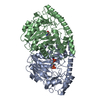


 PDBj
PDBj Assembly
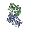

 Mass: 94.971 Da / Num. of mol.: 2 / Source method: obtained synthetically / Formula: PO4
 Mass: 62.068 Da / Num. of mol.: 8 / Source method: obtained synthetically / Formula: C2H6O2
 Mass: 18.015 Da / Num. of mol.: 402 / Source method: isolated from a natural source / Formula: H2O
 Sample preparation
 / Beamline: BL9-2 / Wavelength: 0.91162,0.97932
 Processing