 Movie
Movie Controller

[English] 日本語
 Yorodumi
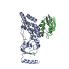
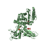
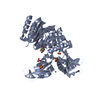
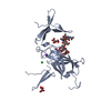
- PDB-3eze: COMPLEX OF THE AMINO TERMINAL DOMAIN OF ENZYME I AND THE HISTIDIN... -
+ Open data

- Basic information
| Entry | Database: PDB / ID: 3eze | ||||||
|---|---|---|---|---|---|---|---|
| Title | COMPLEX OF THE AMINO TERMINAL DOMAIN OF ENZYME I AND THE HISTIDINE-CONTAINING PHOSPHOCARRIER PROTEIN HPR FROM ESCHERICHIA COLI NMR, RESTRAINED REGULARIZED MEAN STRUCTURE | ||||||
 Components Components |
| ||||||
 Keywords Keywords | TRANSFERASE / PHOSPHOTRANSFERASE / KINASE / SUGAR TRANSPORT | ||||||
| Function / homology |  Function and homology information Function and homology informationphosphotransferase activity, nitrogenous group as acceptor / phosphoenolpyruvate-protein phosphotransferase / phosphoenolpyruvate-protein phosphotransferase activity / N-acetylglucosamine transport / antisigma factor binding / regulation of carbon utilization / positive regulation of glycogen catabolic process / phosphoenolpyruvate-dependent sugar phosphotransferase system / enzyme regulator activity / enzyme inhibitor activity ...phosphotransferase activity, nitrogenous group as acceptor / phosphoenolpyruvate-protein phosphotransferase / phosphoenolpyruvate-protein phosphotransferase activity / N-acetylglucosamine transport / antisigma factor binding / regulation of carbon utilization / positive regulation of glycogen catabolic process / phosphoenolpyruvate-dependent sugar phosphotransferase system / enzyme regulator activity / enzyme inhibitor activity / enzyme activator activity / kinase activity / metal ion binding / identical protein binding / cytosol Similarity search - Function | ||||||
| Biological species |  | ||||||
| Method | SOLUTION NMR / simulated annealing | ||||||
 Authors Authors | Clore, G.M. / Garrett, D.S. / Gronenborn, A.M. | ||||||
 Citation Citation | Journal: Nat.Struct.Biol. / Year: 1999 Title: Solution structure of the 40,000 Mr phosphoryl transfer complex between the N-terminal domain of enzyme I and HPr. Authors: Garrett, D.S. / Seok, Y.J. / Peterkofsky, A. / Gronenborn, A.M. / Clore, G.M. #1: Journal: Protein Sci. / Year: 1998Title: Tautomeric State and pKa of the Phosphorylated Active Site Histidine in the N-Terminal Domain of Enzyme I of the Escherichia coli Phosphoenolpyruvate: Sugar Phosphotransferase System Authors: Garrett, D.S. / Seok, Y.J. / Peterkofsky, A. / Clore, G.M. / Gronenborn, A.M. #2: Journal: Biochemistry / Year: 1997Title: Solution Structure of the 30 kDa N-Terminal Domain of Enzyme I of the Escherichia Coli Phosphoenolpyruvate:Sugar Phosphotransferase System by Multidimensional NMR Authors: Garrett, D.S. / Seok, Y.J. / Liao, D.I. / Peterkofsky, A. / Gronenborn, A.M. / Clore, G.M. #3: Journal: Biochemistry / Year: 1997Title: Identification by NMR of the Binding Surface for the Histidine-Containing Phosphocarrier Protein HPr on the N-Terminal Domain of Enzyme I of the Escherichia Coli Phosphotransferase System Authors: Garrett, D.S. / Seok, Y.J. / Peterkofsky, A. / Clore, G.M. / Gronenborn, A.M. | ||||||
| History |
|
- Structure visualization
| Structure viewer | Molecule: MolmilJmol/JSmol |
|---|
- Downloads & links
-Download
| PDBx/mmCIF format | 3eze.cif.gz | 123.4 KB | Display | PDBx/mmCIF format |
|---|---|---|---|---|
| PDB format | pdb3eze.ent.gz | 97.6 KB | Display | PDB format |
| PDBx/mmJSON format | 3eze.json.gz | Tree view | PDBx/mmJSON format | |
| Others |  Other downloads Other downloads |
-Validation report
| Arichive directory | https://data.pdbj.org/pub/pdb/validation_reports/ez/3ezeftp://data.pdbj.org/pub/pdb/validation_reports/ez/3eze | HTTPS FTP |
|---|
-Related structure data


-Links
 PDBj
PDBj
- Assembly
| Deposited unit | 
| |||||||||
|---|---|---|---|---|---|---|---|---|---|---|
| 1 |
| |||||||||
| NMR ensembles |
|
-Components
| #1: Protein | Mass: 28381.316 Da / Num. of mol.: 1 / Fragment: AMINO-TERMINAL DOMAIN RESIDUES 1 - 259 Source method: isolated from a genetically manipulated source Source: (gene. exp.) References: UniProt: P08839, phosphoenolpyruvate-protein phosphotransferase |
|---|---|
| #2: Protein | Mass: 9129.332 Da / Num. of mol.: 1 Source method: isolated from a genetically manipulated source Source: (gene. exp.) |
| #3: Chemical | ChemComp-PO3 /   Mass: 78.972 Da / Num. of mol.: 1 / Source method: obtained synthetically / Formula: PO3 Mass: 78.972 Da / Num. of mol.: 1 / Source method: obtained synthetically / Formula: PO3 |
-Experimental details
-Experiment
| Experiment | Method: SOLUTION NMR |
|---|---|
| NMR details | Text: THE 3D STRUCTURE OF THE EIN-HPR COMPLEX WAS SOLVED BY MULTI HETERONUCLEAR NMR AND IS BASED ON 5475 |
- Sample preparation
| Sample conditions | pH: 7 / Temperature: 313 K |
|---|---|
| Crystal grow | *PLUS Method: other / Details: NMR |
-NMR measurement
| NMR spectrometer |
|
|---|
- Processing
| NMR software |
| |||||||||
|---|---|---|---|---|---|---|---|---|---|---|
| Refinement | Method: simulated annealing / Software ordinal: 1 Details: THE STRUCTURES WERE CALCULATED USING THE SIMULATED ANNEALING PROTOCOL OF NILGES ET AL. (1988) FEBS LETT. 229, 129-136 USING THE PROGRAM CNS MODIFIED TO INCORPORATE COUPLING CONSTANT ...Details: THE STRUCTURES WERE CALCULATED USING THE SIMULATED ANNEALING PROTOCOL OF NILGES ET AL. (1988) FEBS LETT. 229, 129-136 USING THE PROGRAM CNS MODIFIED TO INCORPORATE COUPLING CONSTANT RESTRAINTS (GARRETT ET AL. (1984) J. MAGN. RESON. SERIES B 104, 99-103), CARBON CHEMICAL SHIFT RESTRAINTS, (KUSZEWSKI ET AL. (1995) J. MAGN. RESON. SERIES B 106, 92-96) RESTRAINTS, AND RESIDUAL DIPOLAR COUPLING RESTRAINTS (CLORE ET AL. J. MAGN. RESON 131, 159-162 (1998); J. MAGN 133, 216-221 (1998)). IN THIS ENTRY THE LAST COLUMN REPRESENTS THE AVERAGE RMS DIFFERENCE BETWEEN THE INDIVIDUAL SIMULATED ANNEALING STRUCTURES AND THE MEAN COORDINATE POSITIONS. THE LAST COLUMN IN THE INDIVIDUAL SA STRUCTURES HAS NO MEANING. BEST FITTING TO GENERATE THE AVERAGE STRUCTURE IS WITH RESPECT TO RESIDUES 1 - 250 (RESIDUES 251 - 259 ARE DISORDERED IN SOLUTION) OF ENZYME I AND RESIDUES 301-385 OF HPR. RESIDUES 251-259 ARE OMITTED FROM THE MEAN STRUCTURE | |||||||||
| NMR ensemble | Conformer selection criteria: REGULARIZED MEAN STRUCTURE / Conformers calculated total number: 40 / Conformers submitted total number: 1 |
