 Movie
Movie Controller

[English] 日本語
 Yorodumi
- PDB-3ez4: Crystal structure of 3-methyl-2-oxobutanoate hydroxymethyltransfe... -
+ Open data

- Basic information
| Entry | Database: PDB / ID: 3ez4 | ||||||
|---|---|---|---|---|---|---|---|
| Title | Crystal structure of 3-methyl-2-oxobutanoate hydroxymethyltransferase from Burkholderia pseudomallei | ||||||
 Components Components | 3-methyl-2-oxobutanoate hydroxymethyltransferase | ||||||
 Keywords Keywords | TRANSFERASE / Cytoplasm / Magnesium / Metal-binding / Methyltransferase / Pantothenate biosynthesis / Structural Genomics / Seattle Structural Genomics Center for Infectious Disease / SSGCID | ||||||
| Function / homology |  Function and homology information Function and homology information3-methyl-2-oxobutanoate hydroxymethyltransferase / 3-methyl-2-oxobutanoate hydroxymethyltransferase activity / pantothenate biosynthetic process / magnesium ion binding / cytoplasm Similarity search - Function | ||||||
| Biological species |  Burkholderia pseudomallei (bacteria) Burkholderia pseudomallei (bacteria) | ||||||
| Method |  X-RAY DIFFRACTION / SYNCHROTRON / MOLECULAR REPLACEMENT / molecular replacement / Resolution: 2.1 Å X-RAY DIFFRACTION / SYNCHROTRON / MOLECULAR REPLACEMENT / molecular replacement / Resolution: 2.1 Å | ||||||
 Authors Authors | Seattle Structural Genomics Center for Infectious Disease (SSGCID) | ||||||
 Citation Citation | Journal: To be Published Title: Crystal structure of 3-methyl-2-oxobutanoate hydroxymethyltransferase from Burkholderia pseudomallei Authors: Edwards, T.E. | ||||||
| History |
|
- Structure visualization
| Structure viewer | Molecule: MolmilJmol/JSmol |
|---|
- Downloads & links
-Download
| PDBx/mmCIF format | 3ez4.cif.gz | 471.4 KB | Display | PDBx/mmCIF format |
|---|---|---|---|---|
| PDB format | pdb3ez4.ent.gz | 385.6 KB | Display | PDB format |
| PDBx/mmJSON format | 3ez4.json.gz | Tree view | PDBx/mmJSON format | |
| Others |  Other downloads Other downloads |
-Validation report
| Arichive directory | https://data.pdbj.org/pub/pdb/validation_reports/ez/3ez4ftp://data.pdbj.org/pub/pdb/validation_reports/ez/3ez4 | HTTPS FTP |
|---|
-Related structure data
| Related structure data | 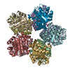 1m3uS S: Starting model for refinement |
|---|---|
| Similar structure data | |
| Other databases |


-Links
 PDBj
PDBj
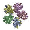
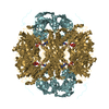
- Assembly
| Deposited unit | 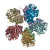
| ||||||||
|---|---|---|---|---|---|---|---|---|---|
| 1 |
| ||||||||
| Unit cell |
|
-Components
| #1: Protein | Mass: 28542.816 Da / Num. of mol.: 10 Source method: isolated from a genetically manipulated source Source: (gene. exp.) Burkholderia pseudomallei (bacteria) / Strain: 1710b / Gene: 721, 76, 808, BPSL2824, panB / Plasmid: BG1861 / Production host: References: UniProt: Q63R49, UniProt: Q3JP15*PLUS, 3-methyl-2-oxobutanoate hydroxymethyltransferase #2: Chemical | ChemComp-TRS /   Mass: 122.143 Da / Num. of mol.: 5 / Source method: obtained synthetically / Formula: C4H12NO3 / Comment: pH buffer*YM Mass: 122.143 Da / Num. of mol.: 5 / Source method: obtained synthetically / Formula: C4H12NO3 / Comment: pH buffer*YM#3: Water | ChemComp-HOH / |  Mass: 18.015 Da / Num. of mol.: 1447 / Source method: isolated from a natural source / Formula: H2O Mass: 18.015 Da / Num. of mol.: 1447 / Source method: isolated from a natural source / Formula: H2O |
|---|
-Experimental details
-Experiment
| Experiment | Method: X-RAY DIFFRACTION / Number of used crystals: 1 |
|---|
- Sample preparation
| Crystal | Density Matthews: 2.38 Å3/Da / Density % sol: 48.4 % |
|---|---|
| Crystal grow | Temperature: 289 K / Method: vapor diffusion, sitting drop / pH: 8.5 Details: PACT screen condition H11, 0.1 M BisTris propane, 20% PEG 3350, 0.2 M Na Citrate, 0.4/0.4 uL drops, 24.4 mg/mL protein, pH 8.5, VAPOR DIFFUSION, SITTING DROP, temperature 289K |
-Data collection
| Diffraction | Mean temperature: 100 K |
|---|---|
| Diffraction source | Source: SYNCHROTRON / Site: APS  / Beamline: 23-ID-D / Wavelength: 0.97934 Å / Beamline: 23-ID-D / Wavelength: 0.97934 Å |
| Detector | Date: Aug 1, 2008 |
| Radiation | Protocol: SINGLE WAVELENGTH / Monochromatic (M) / Laue (L): M / Scattering type: x-ray |
| Radiation wavelength | Wavelength: 0.97934 Å / Relative weight: 1 |
| Reflection | Resolution: 2.1→50 Å / Num. obs: 157415 / % possible obs: 98.5 % / Redundancy: 4.6 % / Rmerge(I) obs: 0.107 / Χ2: 0.924 / Net I/σ(I): 13.461 |
| Reflection shell | Resolution: 2.1→2.18 Å / Redundancy: 4.3 % / Rmerge(I) obs: 0.465 / Mean I/σ(I) obs: 2.3 / Num. unique all: 15125 / Χ2: 0.483 / % possible all: 96 |
-Phasing
| Phasing | Method: molecular replacement |
|---|
- Processing
| Software |
| |||||||||||||||||||||||||||||||||||||||||||||||||||||||||||||||||||||||||||||||||||||
|---|---|---|---|---|---|---|---|---|---|---|---|---|---|---|---|---|---|---|---|---|---|---|---|---|---|---|---|---|---|---|---|---|---|---|---|---|---|---|---|---|---|---|---|---|---|---|---|---|---|---|---|---|---|---|---|---|---|---|---|---|---|---|---|---|---|---|---|---|---|---|---|---|---|---|---|---|---|---|---|---|---|---|---|---|---|---|
| Refinement | Method to determine structure: MOLECULAR REPLACEMENT Starting model: pdb entry 1M3U Resolution: 2.1→38.55 Å / Cor.coef. Fo:Fc: 0.932 / Cor.coef. Fo:Fc free: 0.893 / WRfactor Rfree: 0.228 / WRfactor Rwork: 0.184 / Occupancy max: 1 / Occupancy min: 1 / FOM work R set: 0.831 / SU B: 4.585 / SU ML: 0.121 / SU R Cruickshank DPI: 0.218 / SU Rfree: 0.188 / Cross valid method: THROUGHOUT / σ(F): 0 / ESU R: 0.217 / ESU R Free: 0.188 / Stereochemistry target values: MAXIMUM LIKELIHOOD / Details: HYDROGENS HAVE BEEN ADDED IN THE RIDING POSITIONS
| |||||||||||||||||||||||||||||||||||||||||||||||||||||||||||||||||||||||||||||||||||||
| Solvent computation | Ion probe radii: 0.8 Å / Shrinkage radii: 0.8 Å / VDW probe radii: 1.2 Å / Solvent model: MASK | |||||||||||||||||||||||||||||||||||||||||||||||||||||||||||||||||||||||||||||||||||||
| Displacement parameters | Biso max: 60.39 Å2 / Biso mean: 23.973 Å2 / Biso min: 5.38 Å2
| |||||||||||||||||||||||||||||||||||||||||||||||||||||||||||||||||||||||||||||||||||||
| Refinement step | Cycle: LAST / Resolution: 2.1→38.55 Å
| |||||||||||||||||||||||||||||||||||||||||||||||||||||||||||||||||||||||||||||||||||||
| Refine LS restraints |
| |||||||||||||||||||||||||||||||||||||||||||||||||||||||||||||||||||||||||||||||||||||
| LS refinement shell | Resolution: 2.1→2.152 Å / Total num. of bins used: 20
|
