 Movie
Movie Controller

[English] 日本語
 Yorodumi
- PDB-1glb: STRUCTURE OF THE REGULATORY COMPLEX OF ESCHERICHIA COLI IIIGLC WI... -
+ Open data

- Basic information
| Entry | Database: PDB / ID: 1glb | ||||||
|---|---|---|---|---|---|---|---|
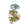
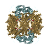
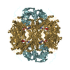
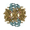
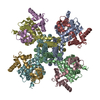
| Title | STRUCTURE OF THE REGULATORY COMPLEX OF ESCHERICHIA COLI IIIGLC WITH GLYCEROL KINASE | ||||||
 Components Components |
| ||||||
 Keywords Keywords | PHOSPHOTRANSFERASE | ||||||
| Function / homology |  Function and homology information Function and homology informationnegative regulation of carbohydrate metabolic process / regulation of carbohydrate utilization / glycerol-3-phosphate metabolic process / negative regulation of maltose transport / enzyme IIA-maltose transporter complex / negative regulation of transmembrane transport / glycerol kinase / glycerol kinase activity / glycerol metabolic process / phosphoenolpyruvate-dependent sugar phosphotransferase system ...negative regulation of carbohydrate metabolic process / regulation of carbohydrate utilization / glycerol-3-phosphate metabolic process / negative regulation of maltose transport / enzyme IIA-maltose transporter complex / negative regulation of transmembrane transport / glycerol kinase / glycerol kinase activity / glycerol metabolic process / phosphoenolpyruvate-dependent sugar phosphotransferase system / transmembrane transporter complex / glycerol catabolic process / kinase activity / DNA damage response / zinc ion binding / ATP binding / membrane / metal ion binding / identical protein binding / cytosol Similarity search - Function | ||||||
| Biological species |  | ||||||
| Method |  X-RAY DIFFRACTION / Resolution: 2.6 Å X-RAY DIFFRACTION / Resolution: 2.6 Å | ||||||
 Authors Authors | Hurley, J.H. / Worthylake, D. / Faber, H.R. / Meadow, N.D. / Roseman, S. / Pettigrew, D.W. / Remington, S.J. | ||||||
 Citation Citation | Journal: Science / Year: 1993 Title: Structure of the regulatory complex of Escherichia coli IIIGlc with glycerol kinase. Authors: Hurley, J.H. / Faber, H.R. / Worthylake, D. / Meadow, N.D. / Roseman, S. / Pettigrew, D.W. / Remington, S.J. | ||||||
| History |
|
- Structure visualization
| Structure viewer | Molecule: MolmilJmol/JSmol |
|---|
- Downloads & links
-Download
| PDBx/mmCIF format | 1glb.cif.gz | 137 KB | Display | PDBx/mmCIF format |
|---|---|---|---|---|
| PDB format | pdb1glb.ent.gz | 105.4 KB | Display | PDB format |
| PDBx/mmJSON format | 1glb.json.gz | Tree view | PDBx/mmJSON format | |
| Others |  Other downloads Other downloads |
-Validation report
| Arichive directory | https://data.pdbj.org/pub/pdb/validation_reports/gl/1glbftp://data.pdbj.org/pub/pdb/validation_reports/gl/1glb | HTTPS FTP |
|---|
-Related structure data


-Links
 PDBj
PDBj- Assembly
| Deposited unit | 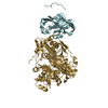
| ||||||||
|---|---|---|---|---|---|---|---|---|---|
| 1 | 
| ||||||||
| Unit cell |
| ||||||||
| Details | GLYCEROL KINASE EXISTS AT PHYSIOLOGICAL CONCENTRATIONS IN AN EQUILIBRIUM BETWEEN FUNCTIONAL DIMERS AND TETRAMERS. THE CRYSTAL CONTAINS TETRAMERS OF GLYCEROL-GLUCOSE-SPECIFIC FACTOR III COMPLEX KINASE WITH EXACT 222 POINT-GROUP SYMMETRY. THE TETRAMER IS LOCATED AT THE INTERSECTION OF THREE TWO-FOLD AXES IN THE CRYSTAL LATTICE. THE FOLLOWING TRANSFORMATIONS WILL PRODUCE A TETRAMER OF THE COMPLEX WHEN APPLIED TO THE COORDINATES IN THIS ENTRY: TRANSFORM 1 (-X, 1-Y, Z) -1.0 0.0 0.0 0.0 0.0 -1.0 0.0 124.90 0.0 0.0 1.0 0.0 TRANSFORM 2 (X, 1-Y, -Z) 1.0 0.0 0.0 0.0 0.0 -1.0 0.0 124.90 0.0 0.0 -1.0 0.0 TRANSFORM 3 (-X, Y, -Z) -1.0 0.0 0.0 0.0 0.0 1.0 0.0 0.0 0.0 0.0 -1.0 0.0 |
-Components
| #1: Protein | Mass: 18141.834 Da / Num. of mol.: 1 Source method: isolated from a genetically manipulated source Source: (gene. exp.) References: UniProt: P69783, protein-Npi-phosphohistidine-sugar phosphotransferase |
|---|---|
| #2: Protein | Mass: 56162.352 Da / Num. of mol.: 1 Source method: isolated from a genetically manipulated source Source: (gene. exp.) |
| #3: Chemical | ChemComp-ADP /   Mass: 427.201 Da / Num. of mol.: 1 / Source method: obtained synthetically / Formula: C10H15N5O10P2 / Comment: ADP, energy-carrying molecule*YM Mass: 427.201 Da / Num. of mol.: 1 / Source method: obtained synthetically / Formula: C10H15N5O10P2 / Comment: ADP, energy-carrying molecule*YM |
| #4: Chemical | ChemComp-GOL /   Mass: 92.094 Da / Num. of mol.: 1 / Source method: obtained synthetically / Formula: C3H8O3 Mass: 92.094 Da / Num. of mol.: 1 / Source method: obtained synthetically / Formula: C3H8O3 |
-Experimental details
-Experiment
| Experiment | Method: X-RAY DIFFRACTION |
|---|
- Sample preparation
| Crystal | Density Matthews: 3.49 Å3/Da / Density % sol: 64.73 % | ||||||||||||||||||||||||||||||
|---|---|---|---|---|---|---|---|---|---|---|---|---|---|---|---|---|---|---|---|---|---|---|---|---|---|---|---|---|---|---|---|
| Crystal grow | *PLUS pH: 6 / Method: vapor diffusion | ||||||||||||||||||||||||||||||
| Components of the solutions | *PLUS
|
-Data collection
| Radiation | Scattering type: x-ray |
|---|---|
| Radiation wavelength | Relative weight: 1 |
| Reflection | *PLUS Highest resolution: 2.6 Å / Num. obs: 24169 / % possible obs: 76 % / Num. measured all: 81994 / Rmerge(I) obs: 0.056 |
| Reflection shell | *PLUS Highest resolution: 2.6 Å / Lowest resolution: 2.7 Å / Num. possible: 58 / Rmerge(I) obs: 0.232 |
- Processing
| Software |
| ||||||||||||||||||||||||||||||||||||||||||||||||||||||||||||
|---|---|---|---|---|---|---|---|---|---|---|---|---|---|---|---|---|---|---|---|---|---|---|---|---|---|---|---|---|---|---|---|---|---|---|---|---|---|---|---|---|---|---|---|---|---|---|---|---|---|---|---|---|---|---|---|---|---|---|---|---|---|
| Refinement | Rfactor Rwork: 0.205 / Rfactor obs: 0.205 / Highest resolution: 2.6 Å | ||||||||||||||||||||||||||||||||||||||||||||||||||||||||||||
| Refinement step | Cycle: LAST / Highest resolution: 2.6 Å
| ||||||||||||||||||||||||||||||||||||||||||||||||||||||||||||
| Refine LS restraints |
|
