 Movie
Movie Controller

[English] 日本語
 Yorodumi
- PDB-1oy0: The crystal Structure of the First Enzyme of Pantothenate Biosynt... -
+ Open data

- Basic information
| Entry | Database: PDB / ID: 1oy0 | ||||||
|---|---|---|---|---|---|---|---|
| Title | The crystal Structure of the First Enzyme of Pantothenate Biosynthetic Pathway, Ketopantoate Hydroxymethyltransferase from Mycobacterium Tuberculosis Shows a Decameric Assembly and Terminal Helix-Swapping | ||||||
 Components Components | Ketopantoate hydroxymethyltransferase | ||||||
 Keywords Keywords | TRANSFERASE / domain swapping / Structural Genomics / PSI / Protein Structure Initiative / TB Structural Genomics Consortium / TBSGC | ||||||
| Function / homology |  Function and homology information Function and homology information3-methyl-2-oxobutanoate hydroxymethyltransferase / 3-methyl-2-oxobutanoate hydroxymethyltransferase activity / pantothenate biosynthetic process / magnesium ion binding / plasma membrane / cytoplasm Similarity search - Function | ||||||
| Biological species |   Mycobacterium tuberculosis (bacteria) Mycobacterium tuberculosis (bacteria) | ||||||
| Method |  X-RAY DIFFRACTION / SAS with averaging / Resolution: 2.8 Å X-RAY DIFFRACTION / SAS with averaging / Resolution: 2.8 Å | ||||||
 Authors Authors | Chaudhuri, B.N. / Sawaya, M.R. / Kim, C.Y. / Waldo, G.S. / Park, M.S. / Terwilliger, T.C. / Yeates, T.O. / TB Structural Genomics Consortium (TBSGC) | ||||||
 Citation Citation | Journal: Structure / Year: 2003 Title: The Crystal Structure of the First Enzyme in the Pantothenate Biosynthetic Pathway, Ketopantoate Hydroxymethyltransferase, from M. tuberculosis Authors: Chaudhuri, B.N. / Sawaya, M.R. / Kim, C.Y. / Waldo, G.S. / Park, M.S. / Terwilliger, T.C. / Yeates, T.O. | ||||||
| History |
|
- Structure visualization
| Structure viewer | Molecule: MolmilJmol/JSmol |
|---|
- Downloads & links
-Download
| PDBx/mmCIF format | 1oy0.cif.gz | 215.6 KB | Display | PDBx/mmCIF format |
|---|---|---|---|---|
| PDB format | pdb1oy0.ent.gz | 177.2 KB | Display | PDB format |
| PDBx/mmJSON format | 1oy0.json.gz | Tree view | PDBx/mmJSON format | |
| Others |  Other downloads Other downloads |
-Validation report
| Arichive directory | https://data.pdbj.org/pub/pdb/validation_reports/oy/1oy0ftp://data.pdbj.org/pub/pdb/validation_reports/oy/1oy0 | HTTPS FTP |
|---|
-Related structure data
| Similar structure data | |
|---|---|
| Other databases |


-Links
 PDBj
PDBj
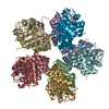
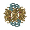
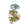
- Assembly
| Deposited unit | 
| ||||||||||
|---|---|---|---|---|---|---|---|---|---|---|---|
| 1 | 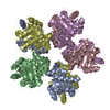
| ||||||||||
| 2 | 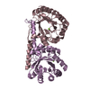
| ||||||||||
| 3 | 
| ||||||||||
| Unit cell |
|
-Components
| #1: Protein | Mass: 29365.287 Da / Num. of mol.: 5 Source method: isolated from a genetically manipulated source Source: (gene. exp.) Mycobacterium tuberculosis (bacteria) / Gene: PANB / Production host: References: UniProt: P0A5Q8, UniProt: P9WIL7*PLUS, 3-methyl-2-oxobutanoate hydroxymethyltransferase #2: Chemical | ChemComp-MG /   Mass: 24.305 Da / Num. of mol.: 5 / Source method: obtained synthetically / Formula: Mg Mass: 24.305 Da / Num. of mol.: 5 / Source method: obtained synthetically / Formula: Mg#3: Water | ChemComp-HOH / |  Mass: 18.015 Da / Num. of mol.: 105 / Source method: isolated from a natural source / Formula: H2O Mass: 18.015 Da / Num. of mol.: 105 / Source method: isolated from a natural source / Formula: H2O |
|---|
-Experimental details
-Experiment
| Experiment | Method: X-RAY DIFFRACTION / Number of used crystals: 1 |
|---|
- Sample preparation
| Crystal | Density Matthews: 2.85 Å3/Da / Density % sol: 56.6 % | ||||||||||||||||||||
|---|---|---|---|---|---|---|---|---|---|---|---|---|---|---|---|---|---|---|---|---|---|
| Crystal grow | Temperature: 298 K / Method: vapor diffusion, hanging drop / pH: 7 Details: Magnesium formate, ethylene glycol, pH 7.0, VAPOR DIFFUSION, HANGING DROP, temperature 298K | ||||||||||||||||||||
| Crystal grow | *PLUS Method: vapor diffusion, hanging drop | ||||||||||||||||||||
| Components of the solutions | *PLUS
|
-Data collection
| Diffraction | Mean temperature: 100 K |
|---|---|
| Diffraction source | Source: ROTATING ANODE / Type: RIGAKU / Wavelength: 1.5418 Å |
| Detector | Type: RIGAKU RAXIS IV / Detector: IMAGE PLATE |
| Radiation | Protocol: SINGLE WAVELENGTH / Monochromatic (M) / Laue (L): M / Scattering type: x-ray |
| Radiation wavelength | Wavelength: 1.5418 Å / Relative weight: 1 |
| Reflection | Resolution: 2.8→50 Å / Num. obs: 36560 / Observed criterion σ(I): -3 / Redundancy: 4.2 % / Biso Wilson estimate: 72.5 Å2 / Rmerge(I) obs: 0.107 / Net I/σ(I): 8.5 |
| Reflection shell | Resolution: 2.8→2.83 Å / Redundancy: 4.3 % / Rmerge(I) obs: 0.468 / Mean I/σ(I) obs: 2.9 |
| Reflection | *PLUS Highest resolution: 2.8 Å / % possible obs: 97.8 % |
| Reflection shell | *PLUS Highest resolution: 2.8 Å / % possible obs: 99.1 % |
- Processing
| Software |
| |||||||||||||||||||||
|---|---|---|---|---|---|---|---|---|---|---|---|---|---|---|---|---|---|---|---|---|---|---|
| Refinement | Method to determine structure: SAS with averaging / Resolution: 2.8→92.53 Å / Rfactor Rfree error: 0.006 / Isotropic thermal model: RESTRAINED / Cross valid method: THROUGHOUT / σ(F): 0 / Stereochemistry target values: Engh & Huber
| |||||||||||||||||||||
| Solvent computation | Solvent model: FLAT MODEL / Bsol: 37.469 Å2 / ksol: 0.348838 e/Å3 | |||||||||||||||||||||
| Displacement parameters | Biso mean: 48.2 Å2
| |||||||||||||||||||||
| Refine analyze | Luzzati coordinate error free: 0.47 Å / Luzzati sigma a free: 0.54 Å | |||||||||||||||||||||
| Refinement step | Cycle: LAST / Resolution: 2.8→92.53 Å
| |||||||||||||||||||||
| Refine LS restraints |
| |||||||||||||||||||||
| Refine LS restraints NCS | NCS model details: CONSTR | |||||||||||||||||||||
| LS refinement shell | Resolution: 2.8→2.98 Å / Rfactor Rfree error: 0.019 / Total num. of bins used: 6
| |||||||||||||||||||||
| Xplor file |
| |||||||||||||||||||||
| Refinement | *PLUS Highest resolution: 2.8 Å / Lowest resolution: 50 Å | |||||||||||||||||||||
| Solvent computation | *PLUS | |||||||||||||||||||||
| Displacement parameters | *PLUS | |||||||||||||||||||||
| Refine LS restraints | *PLUS
| |||||||||||||||||||||
| LS refinement shell | *PLUS Lowest resolution: 2.85 Å / Rfactor Rfree: 0.342 / Rfactor Rwork: 0.313 |
