 Movie
Movie Controller


+ Open data

- Basic information
| Entry | Database: PDB / ID: 3exr | ||||||
|---|---|---|---|---|---|---|---|
| Title | Crystal structure of KGPDC from Streptococcus mutans | ||||||
 Components Components | RmpD (Hexulose-6-phosphate synthase) | ||||||
 Keywords Keywords | LYASE / beta barrel | ||||||
| Function / homology |  Function and homology information Function and homology information3-hexulose-6-phosphate synthase / hexulose-6-phosphate synthase activity / 3-dehydro-L-gulonate-6-phosphate decarboxylase activity / L-ascorbic acid catabolic process / orotidine-5'-phosphate decarboxylase activity / 'de novo' pyrimidine nucleobase biosynthetic process / one-carbon metabolic process / metal ion binding Similarity search - Function | ||||||
| Biological species |  Streptococcus mutans (bacteria) Streptococcus mutans (bacteria) | ||||||
| Method |  X-RAY DIFFRACTION / SYNCHROTRON / MOLECULAR REPLACEMENT / Resolution: 1.7 Å X-RAY DIFFRACTION / SYNCHROTRON / MOLECULAR REPLACEMENT / Resolution: 1.7 Å | ||||||
 Authors Authors | Li, G.L. / Liu, X. / Li, L.F. / Su, X.D. | ||||||
 Citation Citation | Journal: Biochem.Biophys.Res.Commun. / Year: 2009 Title: Open-closed conformational change revealed by the crystal structures of 3-keto-L-gulonate 6-phosphate decarboxylase from Streptococcus mutans Authors: Li, G.L. / Liu, X. / Nan, J. / Brostromer, E. / Li, L.F. / Su, X.D. | ||||||
| History |
|
- Structure visualization
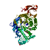
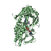
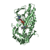
| Structure viewer | Molecule: MolmilJmol/JSmol |
|---|
- Downloads & links
-Download
| PDBx/mmCIF format | 3exr.cif.gz | 345.1 KB | Display | PDBx/mmCIF format |
|---|---|---|---|---|
| PDB format | pdb3exr.ent.gz | 283.6 KB | Display | PDB format |
| PDBx/mmJSON format | 3exr.json.gz | Tree view | PDBx/mmJSON format | |
| Others |  Other downloads Other downloads |
-Validation report
| Arichive directory | https://data.pdbj.org/pub/pdb/validation_reports/ex/3exrftp://data.pdbj.org/pub/pdb/validation_reports/ex/3exr | HTTPS FTP |
|---|
-Related structure data
| Related structure data | 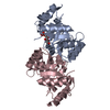 3exsC 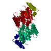 3extSC C: citing same article ( S: Starting model for refinement |
|---|---|
| Similar structure data |


-Links
 PDBj
PDBj
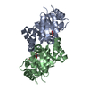
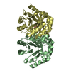
- Assembly
| Deposited unit | 
| ||||||||
|---|---|---|---|---|---|---|---|---|---|
| 1 | 
| ||||||||
| 2 | 
| ||||||||
| Unit cell |
|
-Components
| #1: Protein | Mass: 23850.229 Da / Num. of mol.: 4 Source method: isolated from a genetically manipulated source Source: (gene. exp.) Streptococcus mutans (bacteria) / Gene: ulaD / Plasmid: pET28a / Production host: References: UniProt: Q93DA8, 3-dehydro-L-gulonate-6-phosphate decarboxylase #2: Water | ChemComp-HOH / |  Mass: 18.015 Da / Num. of mol.: 822 / Source method: isolated from a natural source / Formula: H2O Mass: 18.015 Da / Num. of mol.: 822 / Source method: isolated from a natural source / Formula: H2O |
|---|
-Experimental details
-Experiment
| Experiment | Method: X-RAY DIFFRACTION / Number of used crystals: 1 |
|---|
- Sample preparation
| Crystal | Density Matthews: 2.61 Å3/Da / Density % sol: 52.85 % |
|---|---|
| Crystal grow | Temperature: 289 K / Method: vapor diffusion, hanging drop / pH: 7.5 Details: 0.2M KCl, 0.05M HEPES, pH7.5, 35%(v/v) pentaerythritol propoxylate (5/4 PO/OH), VAPOR DIFFUSION, HANGING DROP, temperature 289K |
-Data collection
| Diffraction | Mean temperature: 100 K | ||||||||||||||||||||||||||||||||||||||||||||||||||||||||||||||||||||||
|---|---|---|---|---|---|---|---|---|---|---|---|---|---|---|---|---|---|---|---|---|---|---|---|---|---|---|---|---|---|---|---|---|---|---|---|---|---|---|---|---|---|---|---|---|---|---|---|---|---|---|---|---|---|---|---|---|---|---|---|---|---|---|---|---|---|---|---|---|---|---|---|
| Diffraction source | Source: SYNCHROTRON / Site: MAX II  / Beamline: I711 / Wavelength: 0.9798 Å / Beamline: I711 / Wavelength: 0.9798 Å | ||||||||||||||||||||||||||||||||||||||||||||||||||||||||||||||||||||||
| Detector | Type: MARMOSAIC 225 mm CCD / Detector: CCD / Date: Jul 27, 2006 | ||||||||||||||||||||||||||||||||||||||||||||||||||||||||||||||||||||||
| Radiation | Protocol: SINGLE WAVELENGTH / Monochromatic (M) / Laue (L): M / Scattering type: x-ray | ||||||||||||||||||||||||||||||||||||||||||||||||||||||||||||||||||||||
| Radiation wavelength | Wavelength: 0.9798 Å / Relative weight: 1 | ||||||||||||||||||||||||||||||||||||||||||||||||||||||||||||||||||||||
| Reflection | Resolution: 1.5→50 Å / Num. obs: 158531 / % possible obs: 99.2 % / Observed criterion σ(I): -3 / Biso Wilson estimate: 21.605 Å2 / Rmerge(I) obs: 0.083 | ||||||||||||||||||||||||||||||||||||||||||||||||||||||||||||||||||||||
| Reflection shell |
|
- Processing
| Software |
| ||||||||||||||||||||||||||||||||||||||||||||||||||||||||||||||||||||||||||||||||||||||||||
|---|---|---|---|---|---|---|---|---|---|---|---|---|---|---|---|---|---|---|---|---|---|---|---|---|---|---|---|---|---|---|---|---|---|---|---|---|---|---|---|---|---|---|---|---|---|---|---|---|---|---|---|---|---|---|---|---|---|---|---|---|---|---|---|---|---|---|---|---|---|---|---|---|---|---|---|---|---|---|---|---|---|---|---|---|---|---|---|---|---|---|---|
| Refinement | Method to determine structure: MOLECULAR REPLACEMENT Starting model: PDB ENTRY 3EXT Resolution: 1.7→45.64 Å / Cor.coef. Fo:Fc: 0.929 / Cor.coef. Fo:Fc free: 0.909 / WRfactor Rfree: 0.234 / WRfactor Rwork: 0.196 / Occupancy max: 1 / Occupancy min: 1 / FOM work R set: 0.815 / SU B: 6.388 / SU ML: 0.093 / SU R Cruickshank DPI: 0.154 / SU Rfree: 0.111 / Cross valid method: THROUGHOUT / σ(F): 0 / ESU R: 0.154 / ESU R Free: 0.111 / Stereochemistry target values: MAXIMUM LIKELIHOOD / Details: HYDROGENS HAVE BEEN ADDED IN THE RIDING POSITIONS
| ||||||||||||||||||||||||||||||||||||||||||||||||||||||||||||||||||||||||||||||||||||||||||
| Solvent computation | Ion probe radii: 0.8 Å / Shrinkage radii: 0.8 Å / VDW probe radii: 1.2 Å / Solvent model: MASK | ||||||||||||||||||||||||||||||||||||||||||||||||||||||||||||||||||||||||||||||||||||||||||
| Displacement parameters | Biso max: 45.86 Å2 / Biso mean: 17.915 Å2 / Biso min: 6.48 Å2
| ||||||||||||||||||||||||||||||||||||||||||||||||||||||||||||||||||||||||||||||||||||||||||
| Refinement step | Cycle: LAST / Resolution: 1.7→45.64 Å
| ||||||||||||||||||||||||||||||||||||||||||||||||||||||||||||||||||||||||||||||||||||||||||
| Refine LS restraints |
| ||||||||||||||||||||||||||||||||||||||||||||||||||||||||||||||||||||||||||||||||||||||||||
| LS refinement shell | Resolution: 1.7→1.744 Å / Total num. of bins used: 20
|
