[English] 日本語
Yorodumi
- PDB-3eu9: The ankyrin repeat domain of Huntingtin interacting protein 14 -

+
Open data


ID or keywords:

Loading...

-
Basic information

Entry
Database: PDB / ID: 3eu9
TitleThe ankyrin repeat domain of Huntingtin interacting protein 14
ComponentsHuntingtin-interacting protein 14
Keywordsprotein binding / methyl-lysine-binding protein / epigenetics / ankyrin repeats / methyllyine binding / Huntingtin interacting protein 14 / Acyltransferase / ANK repeat / Cytoplasmic vesicle / Golgi apparatus / Membrane / Metal-binding / Oncogene / Phosphoprotein / Transferase / Transmembrane / Zinc-finger
Function / homology
Function and homology information


regulation of neurotrophin TRK receptor signaling pathway / protein-cysteine S-myristoyltransferase activity / protein-cysteine S-stearoyltransferase activity / protein S-acyltransferase / protein palmitoylation / protein-cysteine S-palmitoyltransferase activity / palmitoyltransferase activity / regulation of programmed cell death / Golgi-associated vesicle membrane / lipoprotein transport ...regulation of neurotrophin TRK receptor signaling pathway / protein-cysteine S-myristoyltransferase activity / protein-cysteine S-stearoyltransferase activity / protein S-acyltransferase / protein palmitoylation / protein-cysteine S-palmitoyltransferase activity / palmitoyltransferase activity / regulation of programmed cell death / Golgi-associated vesicle membrane / lipoprotein transport / Transferases; Acyltransferases; Transferring groups other than aminoacyl groups / axonogenesis / regulation of ERK1 and ERK2 cascade / cell projection / presynaptic membrane / positive regulation of canonical NF-kappaB signal transduction / Golgi membrane / intracellular membrane-bounded organelle / signaling receptor binding / Golgi apparatus / identical protein binding
Similarity search - Function
Palmitoyltransferase, DHHC domain / DHHC palmitoyltransferase / DHHC domain profile. / Ankyrin repeat-containing domain / Ankyrin repeats (3 copies) / Ankyrin repeat profile. / Ankyrin repeat region circular profile. / ankyrin repeats / Ankyrin repeat / Ankyrin repeat-containing domain superfamily ...Palmitoyltransferase, DHHC domain / DHHC palmitoyltransferase / DHHC domain profile. / Ankyrin repeat-containing domain / Ankyrin repeats (3 copies) / Ankyrin repeat profile. / Ankyrin repeat region circular profile. / ankyrin repeats / Ankyrin repeat / Ankyrin repeat-containing domain superfamily / Serine Threonine Protein Phosphatase 5, Tetratricopeptide repeat / Alpha Horseshoe / Mainly Alpha
Similarity search - Domain/homology
HISTIDINE / Palmitoyltransferase ZDHHC17
Similarity search - Component
Biological speciesHomo sapiens (human)
MethodX-RAY DIFFRACTION / SYNCHROTRON / MAD / Resolution: 1.99 Å
AuthorsGao, T. / Collins, R.E. / Horton, J.R. / Zhang, R. / Zhang, X. / Cheng, X.
CitationJournal: Proteins / Year: 2009
Title: The ankyrin repeat domain of Huntingtin interacting protein 14 contains a surface aromatic cage, a potential site for methyl-lysine binding.
Authors: Gao, T. / Collins, R.E. / Horton, J.R. / Zhang, X. / Zhang, R. / Dhayalan, A. / Tamas, R. / Jeltsch, A. / Cheng, X.
History
DepositionOct 9, 2008Deposition site: RCSB / Processing site: RCSB
Revision 1.0Jun 23, 2009Provider: repository / Type: Initial release
Revision 1.1Jul 13, 2011Group: Non-polymer description / Version format compliance
Revision 1.2Dec 27, 2023Group: Data collection / Database references / Derived calculations
Category: chem_comp_atom / chem_comp_bond ...chem_comp_atom / chem_comp_bond / database_2 / struct_site
Item: _database_2.pdbx_DOI / _database_2.pdbx_database_accession ..._database_2.pdbx_DOI / _database_2.pdbx_database_accession / _struct_site.pdbx_auth_asym_id / _struct_site.pdbx_auth_comp_id / _struct_site.pdbx_auth_seq_id

-
Structure visualization

Structure viewerMolecule:
MolmilJmol/JSmol

Downloads & links

-
Assembly

Deposited unit
A: Huntingtin-interacting protein 14
B: Huntingtin-interacting protein 14
C: Huntingtin-interacting protein 14
hetero molecules


Theoretical massNumber of molelcules
Total (without water)81,62113
Polymers80,6083
Non-polymers1,01310
Water6,161342
1
A: Huntingtin-interacting protein 14
B: Huntingtin-interacting protein 14
C: Huntingtin-interacting protein 14
hetero molecules

A: Huntingtin-interacting protein 14
B: Huntingtin-interacting protein 14
C: Huntingtin-interacting protein 14
hetero molecules


Theoretical massNumber of molelcules
Total (without water)163,24226
Polymers161,2176
Non-polymers2,02620
Water1086
TypeNameSymmetry operationNumber
identity operation1_555x,y,z1
crystal symmetry operation10_554-y,-x,-z-1/61
Buried area15010 Å2
ΔGint-224 kcal/mol
Surface area51010 Å2
MethodPISA
2
A: Huntingtin-interacting protein 14
C: Huntingtin-interacting protein 14
hetero molecules


Theoretical massNumber of molelcules
Total (without water)54,56010
Polymers53,7392
Non-polymers8218
Water362
TypeNameSymmetry operationNumber
identity operation1_555x,y,z1
Buried area2890 Å2
ΔGint-73 kcal/mol
Surface area19440 Å2
MethodPISA
Unit cell
Length a, b, c (Å)106.660, 106.660, 255.530
Angle α, β, γ (deg.)90.00, 90.00, 120.00
Int Tables number178
Space group name H-MP6122
Components on special symmetry positions
IDModelComponents
11A-347-

HOH

Details
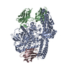
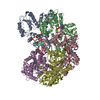
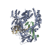
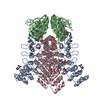
The crystallographic asymmetric unit contains three monomer molecules.

-
Components

#1: Protein Huntingtin-interacting protein 14 / Hip14 / Palmitoyltransferase ZDHHC17 / Zinc finger DHHC domain-containing protein 17 / DHHC-17 / ...Hip14 / Palmitoyltransferase ZDHHC17 / Zinc finger DHHC domain-containing protein 17 / DHHC-17 / Huntingtin-interacting protein 3 / Huntingtin-interacting protein H / Huntingtin yeast partner H / Putative NF-kappa-B-activating protein 205 / Putative MAPK-activating protein PM11


Mass: 26869.482 Da / Num. of mol.: 3 / Fragment: ankyrin repeats
Source method: isolated from a genetically manipulated source
Details: The gene fragment encoding the N-terminal 51-288 residues of HIP14 was amplified from ATCC Clone 5266097 and cloned into the NdeI and XhoI restriction sites of a modified pET28b vector ...Details: The gene fragment encoding the N-terminal 51-288 residues of HIP14 was amplified from ATCC Clone 5266097 and cloned into the NdeI and XhoI restriction sites of a modified pET28b vector containing 6xHis tag followed by SUMO tag, yielding pXC650.
Source: (gene. exp.) Homo sapiens (human) / Gene: ZDHHC17, HIP14, HIP3, HYPH, KIAA0946, HSPC294 / Plasmid: pXC650 / Production host: Escherichia coli (E. coli)
References: UniProt: Q8IUH5, Transferases; Acyltransferases; Transferring groups other than aminoacyl groups
#2: Chemical ChemComp-HIS / HISTIDINE


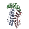
Type: L-peptide linking / Mass: 156.162 Da / Num. of mol.: 1 / Source method: obtained synthetically / Formula: C6H10N3O2
#3: Chemical
ChemComp-SO4 / SULFATE ION


Mass: 96.063 Da / Num. of mol.: 7 / Source method: obtained synthetically / Formula: SO4
#4: Chemical ChemComp-GOL / GLYCEROL / GLYCERIN / PROPANE-1,2,3-TRIOL


Mass: 92.094 Da / Num. of mol.: 2 / Source method: obtained synthetically / Formula: C3H8O3
#5: Water ChemComp-HOH / water


Mass: 18.015 Da / Num. of mol.: 342 / Source method: isolated from a natural source / Formula: H2O

-
Experimental details

-
Experiment

ExperimentMethod: X-RAY DIFFRACTION / Number of used crystals: 2

-
Sample preparation

CrystalDensity Matthews: 2.6 Å3/Da / Density % sol: 52.74 %
Crystal growTemperature: 289 K / pH: 5.5
Details: 100 mM (NH4)2SO4, 25% polyethylene glycol (PEG) 3350, 0.1M Bis-Tris, pH 5.5. , VAPOR DIFFUSION, HANGING DROP, temperature 289K

-
Data collection

Diffraction
IDMean temperature (K)Crystal-ID
11001
21
Diffraction sourceSource: SYNCHROTRON / Site: APS / Beamline: 19-ID / Wavelength: 0.9796, 0.9794
DetectorType: ADSC QUANTUM 315 / Detector: CCD / Date: Dec 10, 2007
Radiation
IDProtocolMonochromatic (M) / Laue (L)Scattering typeWavelength-ID
1MADMx-ray1
2x-ray1
Radiation wavelength
IDWavelength (Å)Relative weight
10.97961
20.97941
ReflectionResolution: 1.99→35 Å / Num. obs: 59687 / % possible obs: 99.1 % / Observed criterion σ(I): -3 / Redundancy: 11 % / Biso Wilson estimate: 31.3 Å2 / Rmerge(I) obs: 0.078 / Net I/σ(I): 14
Reflection shellResolution: 1.99→2.05 Å / Rmerge(I) obs: 0.404 / % possible all: 99.9

-
Processing

Software
NameVersionClassification
HKL-2000data collection
SOLVEphasing
CNS1.1refinement
HKL-2000data reduction
HKL-2000data scaling
RefinementMethod to determine structure: MAD / Resolution: 1.99→34.27 Å / Rfactor Rfree error: 0.005 / Occupancy max: 1 / Occupancy min: 0.5 / Data cutoff high absF: 553241.64 / Data cutoff low absF: 0 / Isotropic thermal model: OVERALL ANISOTROPIC B VALUE / Cross valid method: THROUGHOUT / σ(F): 0 / Stereochemistry target values: ENGH & HUBER
RfactorNum. reflection% reflectionSelection details
Rfree0.251 2937 4.9 %RANDOM
Rwork0.222 ---
obs0.222 58266 97.5 %-
Solvent computationSolvent model: FLAT MODEL / Bsol: 61.8752 Å2 / ksol: 0.4 e/Å3
Displacement parametersBiso mean: 40.06 Å2
Baniso -1Baniso -2Baniso -3
1-0.197 Å20 Å20 Å2
2--0.197 Å20 Å2
3----0.395 Å2
Refine analyze
FreeObs
Luzzati coordinate error0.29 Å0.25 Å
Luzzati d res low-34.3 Å
Luzzati sigma a0.2 Å0.15 Å
Refinement stepCycle: LAST / Resolution: 1.99→34.27 Å
ProteinNucleic acidLigandSolventTotal
Num. atoms5212 0 57 342 5611
Refine LS restraints
Refine-IDTypeDev idealDev ideal target
X-RAY DIFFRACTIONc_bond_d0.005
X-RAY DIFFRACTIONc_bond_d_na
X-RAY DIFFRACTIONc_bond_d_prot
X-RAY DIFFRACTIONc_angle_d
X-RAY DIFFRACTIONc_angle_d_na
X-RAY DIFFRACTIONc_angle_d_prot
X-RAY DIFFRACTIONc_angle_deg1.1
X-RAY DIFFRACTIONc_angle_deg_na
X-RAY DIFFRACTIONc_angle_deg_prot
X-RAY DIFFRACTIONc_dihedral_angle_d20.3
X-RAY DIFFRACTIONc_dihedral_angle_d_na
X-RAY DIFFRACTIONc_dihedral_angle_d_prot
X-RAY DIFFRACTIONc_improper_angle_d0.75
X-RAY DIFFRACTIONc_improper_angle_d_na
X-RAY DIFFRACTIONc_improper_angle_d_prot
X-RAY DIFFRACTIONc_mcbond_it0.9281.5
X-RAY DIFFRACTIONc_mcangle_it1.5212
X-RAY DIFFRACTIONc_scbond_it1.3492
X-RAY DIFFRACTIONc_scangle_it2.0192.5
LS refinement shellResolution: 1.99→2.06 Å / Rfactor Rfree error: 0.017 / Total num. of bins used: 10
RfactorNum. reflection% reflection
Rfree0.289 275 5.2 %
Rwork0.269 4980 -
obs--89.5 %
Xplor fileSerial no: 1 / Param file: protein_rep.param / Topol file: protein.top

+
About Yorodumi

-
News

-
Feb 9, 2022. New format data for meta-information of EMDB entries

New format data for meta-information of EMDB entries

  • Version 3 of the EMDB header file is now the official format.
  • The previous official version 1.9 will be removed from the archive.

Related info.:EMDB header

External links:wwPDB to switch to version 3 of the EMDB data model

-
Aug 12, 2020. Covid-19 info

Covid-19 info

URL: https://pdbjlvh1.pdbj.org/emnavi/covid19.php

New page: Covid-19 featured information page in EM Navigator.

Related info.:Covid-19 info / Mar 5, 2020. Novel coronavirus structure data

+
Mar 5, 2020. Novel coronavirus structure data

Novel coronavirus structure data

Related info.:Yorodumi Speices / Aug 12, 2020. Covid-19 info

External links:COVID-19 featured content - PDBj / Molecule of the Month (242):Coronavirus Proteases

+
Jan 31, 2019. EMDB accession codes are about to change! (news from PDBe EMDB page)

EMDB accession codes are about to change! (news from PDBe EMDB page)

  • The allocation of 4 digits for EMDB accession codes will soon come to an end. Whilst these codes will remain in use, new EMDB accession codes will include an additional digit and will expand incrementally as the available range of codes is exhausted. The current 4-digit format prefixed with “EMD-” (i.e. EMD-XXXX) will advance to a 5-digit format (i.e. EMD-XXXXX), and so on. It is currently estimated that the 4-digit codes will be depleted around Spring 2019, at which point the 5-digit format will come into force.
  • The EM Navigator/Yorodumi systems omit the EMD- prefix.

Related info.:Q: What is EMD? / ID/Accession-code notation in Yorodumi/EM Navigator

External links:EMDB Accession Codes are Changing Soon! / Contact to PDBj

+
Jul 12, 2017. Major update of PDB

Major update of PDB

  • wwPDB released updated PDB data conforming to the new PDBx/mmCIF dictionary.
  • This is a major update changing the version number from 4 to 5, and with Remediation, in which all the entries are updated.
  • In this update, many items about electron microscopy experimental information are reorganized (e.g. em_software).
  • Now, EM Navigator and Yorodumi are based on the updated data.

External links:wwPDB Remediation / Enriched Model Files Conforming to OneDep Data Standards Now Available in the PDB FTP Archive

-
Yorodumi

Thousand views of thousand structures

  • Yorodumi is a browser for structure data from EMDB, PDB, SASBDB, etc.
  • This page is also the successor to EM Navigator detail page, and also detail information page/front-end page for Omokage search.
  • The word "yorodu" (or yorozu) is an old Japanese word meaning "ten thousand". "mi" (miru) is to see.

Related info.:EMDB / PDB / SASBDB / Comparison of 3 databanks / Yorodumi Search / Aug 31, 2016. New EM Navigator & Yorodumi / Yorodumi Papers / Jmol/JSmol / Function and homology information / Changes in new EM Navigator and Yorodumi

Read more