 Movie
Movie Controller


+ Open data

- Basic information
| Entry | Database: PDB / ID: 3esd | ||||||
|---|---|---|---|---|---|---|---|
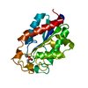
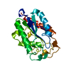
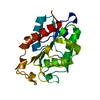
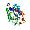
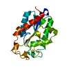
| Title | cut-2b; NCN-Pt-Pincer-Cutinase Hybrid | ||||||
 Components Components | Cutinase 1 | ||||||
 Keywords Keywords | HYDROLASE / protein-metallopincer complex / Glycoprotein / Secreted / Serine esterase | ||||||
| Function / homology |  Function and homology information Function and homology informationcutinase / cutinase activity / carbohydrate catabolic process / extracellular region Similarity search - Function | ||||||
| Biological species |  Fusarium solani f. pisi (fungus) Fusarium solani f. pisi (fungus) | ||||||
| Method |  X-RAY DIFFRACTION / SYNCHROTRON / MOLECULAR REPLACEMENT / Resolution: 1.22 Å X-RAY DIFFRACTION / SYNCHROTRON / MOLECULAR REPLACEMENT / Resolution: 1.22 Å | ||||||
 Authors Authors | Rutten, L. / Mannie, J.P.B.A. / Lutz, M. / Gros, P. | ||||||
 Citation Citation | Journal: Chemistry / Year: 2009 Title: Solid-state structural characterization of cutinase-ECE-pincer-metal hybrids Authors: Rutten, L. / Wieczorek, B. / Mannie, J.P.B.A. / Kruithof, C.A. / Dijkstra, H.P. / Egmond, M.R. / Lutz, M. / Klein Gebbink, R.J.M. / Gros, P. / van Koten, G. | ||||||
| History |
|
- Structure visualization
| Structure viewer | Molecule: MolmilJmol/JSmol |
|---|
- Downloads & links
-Download
| PDBx/mmCIF format | 3esd.cif.gz | 53.1 KB | Display | PDBx/mmCIF format |
|---|---|---|---|---|
| PDB format | pdb3esd.ent.gz | 36.5 KB | Display | PDB format |
| PDBx/mmJSON format | 3esd.json.gz | Tree view | PDBx/mmJSON format | |
| Others |  Other downloads Other downloads |
-Validation report
| Arichive directory | https://data.pdbj.org/pub/pdb/validation_reports/es/3esdftp://data.pdbj.org/pub/pdb/validation_reports/es/3esd | HTTPS FTP |
|---|
-Related structure data
| Related structure data |  3ef3C  3esaC 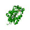 3esbC  3escC  1cuaS S: Starting model for refinement C: citing same article ( |
|---|---|
| Similar structure data |


-Links
 PDBj
PDBj
- Assembly
| Deposited unit | 
| ||||||||
|---|---|---|---|---|---|---|---|---|---|
| 1 |
| ||||||||
| Unit cell |
| ||||||||
| Components on special symmetry positions |
|
-Components
| #1: Protein | Mass: 22294.031 Da / Num. of mol.: 1 / Mutation: N172K Source method: isolated from a genetically manipulated source Source: (gene. exp.) Fusarium solani f. pisi (fungus) / Plasmid: pUC19 / Production host:  |
|---|---|
| #2: Chemical | ChemComp-SXC /   Mass: 654.871 Da / Num. of mol.: 1 / Source method: obtained synthetically / Formula: C21H27BrNO5PPdS2 Mass: 654.871 Da / Num. of mol.: 1 / Source method: obtained synthetically / Formula: C21H27BrNO5PPdS2 |
| #3: Water | ChemComp-HOH /  Mass: 18.015 Da / Num. of mol.: 73 / Source method: isolated from a natural source / Formula: H2O Mass: 18.015 Da / Num. of mol.: 73 / Source method: isolated from a natural source / Formula: H2O |
| Has protein modification | Y |
| Sequence details | SEE REFERENCE 2 IN UNIPROT DATABASE P00590 |
-Experimental details
-Experiment
| Experiment | Method: X-RAY DIFFRACTION / Number of used crystals: 1 |
|---|
- Sample preparation
| Crystal | Density Matthews: 2.5 Å3/Da / Density % sol: 50.87 % |
|---|---|
| Crystal grow | Temperature: 291 K / Method: vapor diffusion / pH: 8.5 Details: 6%(w/v) PEG-3350, 30%(v/v) glycerol, 0.05M BisTrisP, 0.1M potassium thiocyanate, pH 8.5, VAPOR DIFFUSION, temperature 291K |
-Data collection
| Diffraction | Mean temperature: 100 K |
|---|---|
| Diffraction source | Source: SYNCHROTRON / Site: ESRF  / Beamline: ID29 / Wavelength: 1.0332 Å / Beamline: ID29 / Wavelength: 1.0332 Å |
| Detector | Type: ADSC QUANTUM 315 / Detector: CCD / Date: Mar 11, 2007 |
| Radiation | Protocol: SINGLE WAVELENGTH / Monochromatic (M) / Laue (L): M / Scattering type: x-ray |
| Radiation wavelength | Wavelength: 1.0332 Å / Relative weight: 1 |
| Reflection | Resolution: 1.22→41 Å / Num. obs: 61949 / % possible obs: 97.1 % / Observed criterion σ(F): 0 / Observed criterion σ(I): 0 / Redundancy: 3.6 % / Rmerge(I) obs: 0.1 / Net I/σ(I): 5.7 |
| Reflection shell | Resolution: 1.22→1.29 Å / Redundancy: 2.6 % / Rmerge(I) obs: 0.805 / Mean I/σ(I) obs: 1.2 / Num. unique all: 7679 / % possible all: 100 |
- Processing
| Software |
| ||||||||||||||||||||
|---|---|---|---|---|---|---|---|---|---|---|---|---|---|---|---|---|---|---|---|---|---|
| Refinement | Method to determine structure: MOLECULAR REPLACEMENT Starting model: 1CUA Resolution: 1.22→41 Å / σ(F): 0 / σ(I): 0 Details: Twinning was observed in the present crystal and refined with SHELXL using the TWIN instruction, resulting in a twin fraction of 0.5, operator h,-h-k,-l. The point group 3 of the crystal ...Details: Twinning was observed in the present crystal and refined with SHELXL using the TWIN instruction, resulting in a twin fraction of 0.5, operator h,-h-k,-l. The point group 3 of the crystal lattice can be obtained via a coset decomposition of point group 32 of the twin lattice. The twofold twin axis cannot be a real crystallographic operation because this would result in only half a molecule in the asymmetric unit.
| ||||||||||||||||||||
| Refinement step | Cycle: LAST / Resolution: 1.22→41 Å
| ||||||||||||||||||||
| Refine LS restraints |
|
