 Movie
Movie Controller

+ Open data

- Basic information
| Entry | Database: PDB / ID: 3ep1 | ||||||
|---|---|---|---|---|---|---|---|
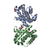
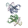
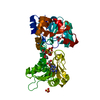
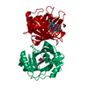
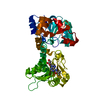
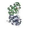
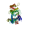
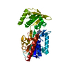
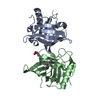
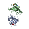
| Title | Structure of the PGRP-Hd from Alvinella pompejana | ||||||
 Components Components | PGRP-Hd - Peptidoglycan recognition protein homologue | ||||||
 Keywords Keywords | IMMUNE SYSTEM / PGRP-Hd / Alvinella pompejana / thermophile / model system | ||||||
| Function / homology |  Function and homology information Function and homology informationN-acetylmuramoyl-L-alanine amidase activity / peptidoglycan catabolic process / zinc ion binding Similarity search - Function | ||||||
| Biological species |  Alvinella pompejana (invertebrata) Alvinella pompejana (invertebrata) | ||||||
| Method |  X-RAY DIFFRACTION / SYNCHROTRON / MOLECULAR REPLACEMENT / Resolution: 2.1 Å X-RAY DIFFRACTION / SYNCHROTRON / MOLECULAR REPLACEMENT / Resolution: 2.1 Å | ||||||
 Authors Authors | Delfosse, V. / Gagniere, N. / Perrodou, E. / Poch, O. / Lecompte, O. / Mayer, C. | ||||||
 Citation Citation | Journal: To be Published Title: Structure of the PGRP-Hd from Alvinella pompejana Authors: Delfosse, V. / Gagniere, N. / Perrodou, E. / Poch, O. / Lecompte, O. / Mayer, C. | ||||||
| History |
|
- Structure visualization
| Structure viewer | Molecule: MolmilJmol/JSmol |
|---|
- Downloads & links
-Download
| PDBx/mmCIF format | 3ep1.cif.gz | 78.1 KB | Display | PDBx/mmCIF format |
|---|---|---|---|---|
| PDB format | pdb3ep1.ent.gz | 58.1 KB | Display | PDB format |
| PDBx/mmJSON format | 3ep1.json.gz | Tree view | PDBx/mmJSON format | |
| Others |  Other downloads Other downloads |
-Validation report
| Arichive directory | https://data.pdbj.org/pub/pdb/validation_reports/ep/3ep1ftp://data.pdbj.org/pub/pdb/validation_reports/ep/3ep1 | HTTPS FTP |
|---|
-Related structure data
| Related structure data |  1yckS S: Starting model for refinement |
|---|---|
| Similar structure data |
-Links
 PDBj
PDBj- Assembly
| Deposited unit | 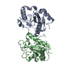
| ||||||||
|---|---|---|---|---|---|---|---|---|---|
| 1 |
| ||||||||
| Unit cell |
|
-Components
| #1: Protein | Mass: 19251.617 Da / Num. of mol.: 2 Source method: isolated from a genetically manipulated source Source: (gene. exp.) Alvinella pompejana (invertebrata) / Gene: PGRP-Hd / Plasmid details: pHGW is based on the pET22b vector / Plasmid: pHGW / Production host:   PDB-3EPI, UniProt: D0VX04*PLUS PDB-3EPI, UniProt: D0VX04*PLUS#2: Water | ChemComp-HOH / |  Mass: 18.015 Da / Num. of mol.: 233 / Source method: isolated from a natural source / Formula: H2O Mass: 18.015 Da / Num. of mol.: 233 / Source method: isolated from a natural source / Formula: H2OHas protein modification | Y | Sequence details | A SEQUENCE DATABASE REFERENCE FOR THIS PROTEIN DOES NOT CURRENTLY EXIST. THIS SEQUENCE WILL BE ...A SEQUENCE DATABASE REFERENCE FOR THIS PROTEIN DOES NOT CURRENTLY EXIST. THIS SEQUENCE WILL BE DEPOSITED IN THE SEQUENCE DATABASE. | |
|---|
-Experimental details
-Experiment
| Experiment | Method: X-RAY DIFFRACTION / Number of used crystals: 1 |
|---|
- Sample preparation
| Crystal | Density Matthews: 2.34 Å3/Da / Density % sol: 47.45 % |
|---|---|
| Crystal grow | Temperature: 291 K / Method: vapor diffusion, hanging drop / pH: 7.4 Details: 200mM Sodium Tartrate, 20% PEG3350, pH7.4, VAPOR DIFFUSION, HANGING DROP, temperature 291K |
-Data collection
| Diffraction | Mean temperature: 100 K |
|---|---|
| Diffraction source | Source: SYNCHROTRON / Site: ESRF  / Beamline: ID23-2 / Wavelength: 0.873 Å / Beamline: ID23-2 / Wavelength: 0.873 Å |
| Detector | Type: MARMOSAIC 225 mm CCD / Detector: CCD / Date: Jun 13, 2008 |
| Radiation | Monochromator: Si 111 / Protocol: SINGLE WAVELENGTH / Monochromatic (M) / Laue (L): M / Scattering type: x-ray |
| Radiation wavelength | Wavelength: 0.873 Å / Relative weight: 1 |
| Reflection | Resolution: 2.1→29 Å / Num. obs: 21905 / % possible obs: 98.5 % / Observed criterion σ(F): 0 / Observed criterion σ(I): 0 / Redundancy: 7.1 % / Biso Wilson estimate: 11.7 Å2 / Rsym value: 0.083 / Net I/σ(I): 15.7 |
| Reflection shell | Resolution: 2.1→2.26 Å / Redundancy: 7.3 % / Mean I/σ(I) obs: 5.7 / Num. unique all: 4084 / Rsym value: 0.37 / % possible all: 96.7 |
- Processing
| Software |
| |||||||||||||||||||||||||
|---|---|---|---|---|---|---|---|---|---|---|---|---|---|---|---|---|---|---|---|---|---|---|---|---|---|---|
| Refinement | Method to determine structure: MOLECULAR REPLACEMENT Starting model: PDB ENTRY 1YCK Resolution: 2.1→28.96 Å / Isotropic thermal model: Restrained / σ(F): 0 / σ(I): 0 / Stereochemistry target values: Engh & Huber
| |||||||||||||||||||||||||
| Displacement parameters | Biso mean: 28.8 Å2
| |||||||||||||||||||||||||
| Refine analyze |
| |||||||||||||||||||||||||
| Refinement step | Cycle: LAST / Resolution: 2.1→28.96 Å
| |||||||||||||||||||||||||
| Refine LS restraints |
| |||||||||||||||||||||||||
| LS refinement shell | Resolution: 2.1→2.23 Å / Rfactor Rfree error: 0.029
|
