 Movie
Movie Controller

[English] 日本語
 Yorodumi
- PDB-3enu: Crystal structure of Nitrollin, a betagamma-crystallin from Nitro... -
+ Open data

- Basic information
| Entry | Database: PDB / ID: 3enu | ||||||
|---|---|---|---|---|---|---|---|
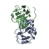
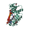
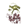
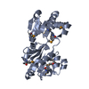
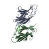
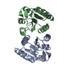
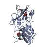
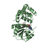
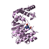
| Title | Crystal structure of Nitrollin, a betagamma-crystallin from Nitrosospira multiformis | ||||||
 Components Components | Putative uncharacterized protein | ||||||
 Keywords Keywords | STRUCTURAL PROTEIN / betagamma crystallin | ||||||
| Function / homology |  Function and homology information Function and homology information | ||||||
| Biological species |  Nitrosospira multiformis (bacteria) Nitrosospira multiformis (bacteria) | ||||||
| Method |  X-RAY DIFFRACTION / MIRAS / Resolution: 1.86 Å X-RAY DIFFRACTION / MIRAS / Resolution: 1.86 Å | ||||||
 Authors Authors | Aravind, P. / Sankaranarayanan, R. | ||||||
 Citation Citation | Journal: J.Mol.Biol. / Year: 2009 Title: Three-dimensional domain swapping in nitrollin, a single-domain betagamma-crystallin from Nitrosospira multiformis, controls protein conformation and stability but not dimerization Authors: Aravind, P. / Suman, S.K. / Mishra, A. / Sharma, Y. / Sankaranarayanan, R. | ||||||
| History |
|
- Structure visualization
| Structure viewer | Molecule: MolmilJmol/JSmol |
|---|
- Downloads & links
-Download
| PDBx/mmCIF format | 3enu.cif.gz | 38.9 KB | Display | PDBx/mmCIF format |
|---|---|---|---|---|
| PDB format | pdb3enu.ent.gz | 25.2 KB | Display | PDB format |
| PDBx/mmJSON format | 3enu.json.gz | Tree view | PDBx/mmJSON format | |
| Others |  Other downloads Other downloads |
-Validation report
| Arichive directory | https://data.pdbj.org/pub/pdb/validation_reports/en/3enuftp://data.pdbj.org/pub/pdb/validation_reports/en/3enu | HTTPS FTP |
|---|
-Related structure data


-Links
 PDBj
PDBj
- Assembly
| Deposited unit | 
| ||||||||
|---|---|---|---|---|---|---|---|---|---|
| 1 | 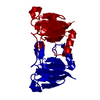
| ||||||||
| Unit cell |
|
-Components
| #1: Protein | Mass: 12949.712 Da / Num. of mol.: 1 Fragment: single domain betagamma-crystallin, residues 27-140 Source method: isolated from a genetically manipulated source Source: (gene. exp.) Nitrosospira multiformis (bacteria) / Strain: ATCC 25196 / Gene: 3786576 / Plasmid: pET21a / Production host: |
|---|---|
| #2: Water | ChemComp-HOH /  Mass: 18.015 Da / Num. of mol.: 125 / Source method: isolated from a natural source / Formula: H2O Mass: 18.015 Da / Num. of mol.: 125 / Source method: isolated from a natural source / Formula: H2O |
| Has protein modification | Y |
-Experimental details
-Experiment
| Experiment | Method: X-RAY DIFFRACTION / Number of used crystals: 1 |
|---|
- Sample preparation
| Crystal | Density Matthews: 2.32 Å3/Da / Density % sol: 47.02 % / Mosaicity: 0.486 ° |
|---|---|
| Crystal grow | Temperature: 277 K / Method: vapor diffusion, sitting drop Details: 20% PEG3350, 0.2M Naformate, vapor diffusion, sitting drop, temperature 277K |
-Data collection
| Diffraction | Mean temperature: 100 K | ||||||||||||||||||||||||||||||||||||||||||||||||||||||||||||||||||
|---|---|---|---|---|---|---|---|---|---|---|---|---|---|---|---|---|---|---|---|---|---|---|---|---|---|---|---|---|---|---|---|---|---|---|---|---|---|---|---|---|---|---|---|---|---|---|---|---|---|---|---|---|---|---|---|---|---|---|---|---|---|---|---|---|---|---|---|
| Diffraction source | Source: ROTATING ANODE / Type: RIGAKU RUH3R / Wavelength: 1.5418 Å | ||||||||||||||||||||||||||||||||||||||||||||||||||||||||||||||||||
| Detector | Type: MAR 345dtb / Detector: IMAGE PLATE / Date: Oct 4, 2007 | ||||||||||||||||||||||||||||||||||||||||||||||||||||||||||||||||||
| Radiation | Protocol: SINGLE WAVELENGTH / Monochromatic (M) / Laue (L): M / Scattering type: x-ray | ||||||||||||||||||||||||||||||||||||||||||||||||||||||||||||||||||
| Radiation wavelength | Wavelength: 1.5418 Å / Relative weight: 1 | ||||||||||||||||||||||||||||||||||||||||||||||||||||||||||||||||||
| Reflection | Resolution: 1.86→25 Å / Num. obs: 10654 / % possible obs: 99.8 % / Redundancy: 6.6 % / Rmerge(I) obs: 0.046 / Χ2: 1.109 / Net I/σ(I): 40.15 | ||||||||||||||||||||||||||||||||||||||||||||||||||||||||||||||||||
| Reflection shell |
|
-Phasing
| Phasing | Method: MIRAS | ||||||||||||||||||||||||||||||||||||||||||||||||
|---|---|---|---|---|---|---|---|---|---|---|---|---|---|---|---|---|---|---|---|---|---|---|---|---|---|---|---|---|---|---|---|---|---|---|---|---|---|---|---|---|---|---|---|---|---|---|---|---|---|
| Phasing MIR | Resolution: 2.4→20 Å / FOM: 0.51 / Reflection: 5062 | ||||||||||||||||||||||||||||||||||||||||||||||||
| Phasing MIR der |
| ||||||||||||||||||||||||||||||||||||||||||||||||
| Phasing MIR der site |
| ||||||||||||||||||||||||||||||||||||||||||||||||
| Phasing MIR shell |
|
- Processing
| Software |
| ||||||||||||||||||||||||||||
|---|---|---|---|---|---|---|---|---|---|---|---|---|---|---|---|---|---|---|---|---|---|---|---|---|---|---|---|---|---|
| Refinement | Method to determine structure: MIRAS / Resolution: 1.86→25 Å / Occupancy max: 1 / Occupancy min: 1 / FOM work R set: 0.874 / σ(F): 0
| ||||||||||||||||||||||||||||
| Solvent computation | Bsol: 42.54 Å2 | ||||||||||||||||||||||||||||
| Displacement parameters | Biso max: 44.65 Å2 / Biso mean: 11.501 Å2 / Biso min: 3 Å2
| ||||||||||||||||||||||||||||
| Refinement step | Cycle: LAST / Resolution: 1.86→25 Å
| ||||||||||||||||||||||||||||
| Refine LS restraints |
| ||||||||||||||||||||||||||||
| Xplor file |
|
