 Movie
Movie Controller

[English] 日本語
 Yorodumi
- PDB-3ela: Crystal structure of active site inhibited coagulation factor VII... -
+ Open data

- Basic information
| Entry | Database: PDB / ID: 3ela | ||||||
|---|---|---|---|---|---|---|---|
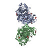
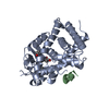
| Title | Crystal structure of active site inhibited coagulation factor VIIA mutant in complex with soluble tissue factor | ||||||
 Components Components |
| ||||||
 Keywords Keywords | HYDROLASE/HYDROLASE INHIBITOR / Serine protease / BLOOD CLOTTING / HYDROLASE-HYDROLASE INHIBITOR complex | ||||||
| Function / homology |  Function and homology information Function and homology informationactivation of plasma proteins involved in acute inflammatory response / activation of blood coagulation via clotting cascade / coagulation factor VIIa / response to Thyroid stimulating hormone / response to astaxanthin / response to thyrotropin-releasing hormone / response to 2,3,7,8-tetrachlorodibenzodioxine / response to carbon dioxide / serine-type peptidase complex / response to genistein ...activation of plasma proteins involved in acute inflammatory response / activation of blood coagulation via clotting cascade / coagulation factor VIIa / response to Thyroid stimulating hormone / response to astaxanthin / response to thyrotropin-releasing hormone / response to 2,3,7,8-tetrachlorodibenzodioxine / response to carbon dioxide / serine-type peptidase complex / response to genistein / response to vitamin K / positive regulation of platelet-derived growth factor receptor signaling pathway / positive regulation of leukocyte chemotaxis / response to thyroxine / NGF-stimulated transcription / response to cholesterol / cytokine receptor activity / positive regulation of positive chemotaxis / : / response to growth hormone / positive regulation of blood coagulation / animal organ regeneration / positive regulation of endothelial cell apoptotic process / positive regulation of TOR signaling / Transport of gamma-carboxylated protein precursors from the endoplasmic reticulum to the Golgi apparatus / Gamma-carboxylation of protein precursors / Removal of aminoterminal propeptides from gamma-carboxylated proteins / positive regulation of endothelial cell proliferation / BMAL1:CLOCK,NPAS2 activates circadian expression / serine-type peptidase activity / positive regulation of interleukin-8 production / circadian rhythm / protein processing / phospholipid binding / response to estrogen / Golgi lumen / cytokine-mediated signaling pathway / positive regulation of angiogenesis / blood coagulation / response to estradiol / extracellular matrix / protease binding / vesicle / response to hypoxia / positive regulation of cell migration / endoplasmic reticulum lumen / serine-type endopeptidase activity / signaling receptor binding / external side of plasma membrane / calcium ion binding / positive regulation of gene expression / cell surface / : / extracellular region / membrane / plasma membrane Similarity search - Function | ||||||
| Biological species |  Homo sapiens (human) Homo sapiens (human) | ||||||
| Method |  X-RAY DIFFRACTION / SYNCHROTRON / MOLECULAR REPLACEMENT / Resolution: 2.2 Å X-RAY DIFFRACTION / SYNCHROTRON / MOLECULAR REPLACEMENT / Resolution: 2.2 Å | ||||||
 Authors Authors | Bjelke, J.R. / Fodje, M. / Svensson, L.A. | ||||||
 Citation Citation | Journal: J.Biol.Chem. / Year: 2008 Title: Mechanism of the Ca2+-induced enhancement of the intrinsic factor VIIa activity Authors: Bjelke, J.R. / Olsen, O.H. / Fodje, M. / Svensson, L.A. / Bang, S. / Bolt, G. / Kragelund, B.B. / Persson, E. | ||||||
| History |
|
- Structure visualization
| Structure viewer | Molecule: MolmilJmol/JSmol |
|---|
- Downloads & links
-Download
| PDBx/mmCIF format | 3ela.cif.gz | 129.8 KB | Display | PDBx/mmCIF format |
|---|---|---|---|---|
| PDB format | pdb3ela.ent.gz | 96.8 KB | Display | PDB format |
| PDBx/mmJSON format | 3ela.json.gz | Tree view | PDBx/mmJSON format | |
| Others |  Other downloads Other downloads |
-Validation report
| Arichive directory | https://data.pdbj.org/pub/pdb/validation_reports/el/3elaftp://data.pdbj.org/pub/pdb/validation_reports/el/3ela | HTTPS FTP |
|---|
-Related structure data
| Related structure data |  1danS S: Starting model for refinement |
|---|---|
| Similar structure data |


-Links
 PDBj
PDBj


- Assembly
| Deposited unit | 
| ||||||||
|---|---|---|---|---|---|---|---|---|---|
| 1 |
| ||||||||
| Unit cell |
|
-Components
-Coagulation factor VIIA ... , 2 types, 2 molecules LH
| #1: Protein | Mass: 17046.975 Da / Num. of mol.: 1 / Fragment: UNP residues 61-212 Source method: isolated from a genetically manipulated source Source: (gene. exp.) Homo sapiens (human) / Gene: F7 / Plasmid: PEE-ACEDELTA36NJ / Cell (production host): CHO-K1 / Production host:   Cricetulus griseus (Chinese hamster) / References: UniProt: P08709, coagulation factor VIIa Cricetulus griseus (Chinese hamster) / References: UniProt: P08709, coagulation factor VIIa |
|---|---|
| #2: Protein | Mass: 28086.164 Da / Num. of mol.: 1 / Fragment: UNP residues 213-466 / Mutation: V158D,E296V,M298Q Source method: isolated from a genetically manipulated source Source: (gene. exp.) Homo sapiens (human) / Gene: F7 / Plasmid: PEE-ACEDELTA36NJ / Cell (production host): CHO-K1 / Production host: Cricetulus griseus (Chinese hamster) / References: UniProt: P08709, coagulation factor VIIa |
-Protein , 1 types, 1 molecules T
| #3: Protein | Mass: 23632.195 Da / Num. of mol.: 1 / Fragment: UNP residues 33-241 Source method: isolated from a genetically manipulated source Source: (gene. exp.) Homo sapiens (human) / Gene: F3 / Plasmid: pET_TF1-209 / Production host:  |
|---|
-Sugars , 2 types, 2 molecules 

| #4: Sugar | ChemComp-GLC /  Type: D-saccharide, alpha linking / Mass: 180.156 Da / Num. of mol.: 1 Type: D-saccharide, alpha linking / Mass: 180.156 Da / Num. of mol.: 1Source method: isolated from a genetically manipulated source Formula: C6H12O6 |
|---|---|
| #5: Sugar | ChemComp-FUC /  Type: L-saccharide, alpha linking / Mass: 164.156 Da / Num. of mol.: 1 Type: L-saccharide, alpha linking / Mass: 164.156 Da / Num. of mol.: 1Source method: isolated from a genetically manipulated source Formula: C6H12O5 |
-Non-polymers , 3 types, 216 molecules 

| #6: Chemical | ChemComp-CA /  Mass: 40.078 Da / Num. of mol.: 1 / Source method: obtained synthetically / Formula: Ca Mass: 40.078 Da / Num. of mol.: 1 / Source method: obtained synthetically / Formula: Ca |
|---|---|
| #7: Chemical | ChemComp-0Z6 /  Type: peptide-like, Peptide-like / Class: Inhibitor / Mass: 504.045 Da / Num. of mol.: 1 / Source method: obtained synthetically / Formula: C25H36ClN6O3 / References: D-Phe-Phe-Arg Chloromethylketone Type: peptide-like, Peptide-like / Class: Inhibitor / Mass: 504.045 Da / Num. of mol.: 1 / Source method: obtained synthetically / Formula: C25H36ClN6O3 / References: D-Phe-Phe-Arg Chloromethylketone |
| #8: Water | ChemComp-HOH / Mass: 18.015 Da / Num. of mol.: 214 / Source method: isolated from a natural source / Formula: H2O |
-Details
| Has protein modification | Y |
|---|---|
| Nonpolymer details | THE UNBOUND FORM OF THE INHIBITOR IS D-PHE-PHE-ARG-CHLOROMETHYLKETONE. UPON REACTION WITH PROTEIN ...THE UNBOUND FORM OF THE INHIBITOR IS D-PHE-PHE-ARG-CHLOROMETH |
-Experimental details
-Experiment
| Experiment | Method: X-RAY DIFFRACTION / Number of used crystals: 1 |
|---|
- Sample preparation
| Crystal | Density Matthews: 3.06 Å3/Da / Density % sol: 59.77 % |
|---|---|
| Crystal grow | Temperature: 298 K / Method: vapor diffusion, hanging drop / pH: 5.6 Details: 100mM NaCitrate, 16%(w/v) PEG4000, 12%(v/v) 1-propanol, pH 5.6, VAPOR DIFFUSION, HANGING DROP, temperature 298K |
-Data collection
| Diffraction | Mean temperature: 80 K |
|---|---|
| Diffraction source | Source: SYNCHROTRON / Site: MAX II  / Beamline: I911-3 / Wavelength: 1.08 Å / Beamline: I911-3 / Wavelength: 1.08 Å |
| Detector | Detector: CCD |
| Radiation | Monochromator: Si(111) / Protocol: SINGLE WAVELENGTH / Monochromatic (M) / Laue (L): M / Scattering type: x-ray |
| Radiation wavelength | Wavelength: 1.08 Å / Relative weight: 1 |
| Reflection | Resolution: 2.2→29.19 Å / Num. all: 34658 / Num. obs: 34658 / % possible obs: 80.3 % / Redundancy: 1.8 % / Biso Wilson estimate: 23.5 Å2 / Rmerge(I) obs: 0.096 |
| Reflection shell | Resolution: 2.2→2.3 Å / Redundancy: 1.3 % / Rmerge(I) obs: 0.229 / Mean I/σ(I) obs: 2.12 / Num. unique all: 1526 / % possible all: 26.1 |
- Processing
| Software |
| |||||||||||||||||||||||||||||||||||||||||||||||||||||||||||||||||||||||||||||||||||||||||||||||
|---|---|---|---|---|---|---|---|---|---|---|---|---|---|---|---|---|---|---|---|---|---|---|---|---|---|---|---|---|---|---|---|---|---|---|---|---|---|---|---|---|---|---|---|---|---|---|---|---|---|---|---|---|---|---|---|---|---|---|---|---|---|---|---|---|---|---|---|---|---|---|---|---|---|---|---|---|---|---|---|---|---|---|---|---|---|---|---|---|---|---|---|---|---|---|---|---|
| Refinement | Method to determine structure: MOLECULAR REPLACEMENT Starting model: PDB ENTRY 1DAN Resolution: 2.2→29.19 Å / Cor.coef. Fo:Fc: 0.909 / Cor.coef. Fo:Fc free: 0.839 / SU B: 10.241 / SU ML: 0.237 / Cross valid method: THROUGHOUT / ESU R: 0.283 / ESU R Free: 0.242 / Stereochemistry target values: MAXIMUM LIKELIHOOD / Details: HYDROGENS HAVE BEEN ADDED IN THE RIDING POSITIONS
| |||||||||||||||||||||||||||||||||||||||||||||||||||||||||||||||||||||||||||||||||||||||||||||||
| Solvent computation | Ion probe radii: 0.8 Å / Shrinkage radii: 0.8 Å / VDW probe radii: 1.2 Å / Solvent model: MASK | |||||||||||||||||||||||||||||||||||||||||||||||||||||||||||||||||||||||||||||||||||||||||||||||
| Displacement parameters | Biso mean: 33.911 Å2
| |||||||||||||||||||||||||||||||||||||||||||||||||||||||||||||||||||||||||||||||||||||||||||||||
| Refinement step | Cycle: LAST / Resolution: 2.2→29.19 Å
| |||||||||||||||||||||||||||||||||||||||||||||||||||||||||||||||||||||||||||||||||||||||||||||||
| Refine LS restraints |
| |||||||||||||||||||||||||||||||||||||||||||||||||||||||||||||||||||||||||||||||||||||||||||||||
| LS refinement shell | Resolution: 2.202→2.259 Å / Total num. of bins used: 20
|
