 Movie
Movie Controller

[English] 日本語
 Yorodumi
- PDB-3ejx: Crystal structure of diaminopimelate epimerase from Arabidopsis t... -
+ Open data

- Basic information
| Entry | Database: PDB / ID: 3ejx | ||||||
|---|---|---|---|---|---|---|---|
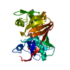
| Title | Crystal structure of diaminopimelate epimerase from Arabidopsis thaliana in complex with LL-AziDAP | ||||||
 Components Components | Diaminopimelate epimerase, chloroplastic | ||||||
 Keywords Keywords | ISOMERASE / Diaminopimelate epimerase / Arabidopsis / PLP-independenet amino acid racemase / aziridino-diaminopimelate | ||||||
| Function / homology |  Function and homology information Function and homology informationdiaminopimelate epimerase / diaminopimelate epimerase activity / : / chloroplast stroma / chloroplast / cytosol Similarity search - Function | ||||||
| Biological species |  | ||||||
| Method |  X-RAY DIFFRACTION / SYNCHROTRON / MOLECULAR REPLACEMENT / Resolution: 1.95 Å X-RAY DIFFRACTION / SYNCHROTRON / MOLECULAR REPLACEMENT / Resolution: 1.95 Å | ||||||
 Authors Authors | Pillai, B. / Moorthie, V.A. / Cherney, M.M. / Vederas, J.C. / James, M.N.G. | ||||||
 Citation Citation | Journal: J.Mol.Biol. / Year: 2009 Title: Crystal structure of diaminopimelate epimerase from Arabidopsis thaliana, an amino acid racemase critical for L-lysine biosynthesis. Authors: Pillai, B. / Moorthie, V.A. / van Belkum, M.J. / Marcus, S.L. / Cherney, M.M. / Diaper, C.M. / Vederas, J.C. / James, M.N. | ||||||
| History |
|
- Structure visualization
| Structure viewer | Molecule: MolmilJmol/JSmol |
|---|
- Downloads & links
-Download
| PDBx/mmCIF format | 3ejx.cif.gz | 376.1 KB | Display | PDBx/mmCIF format |
|---|---|---|---|---|
| PDB format | pdb3ejx.ent.gz | 301.6 KB | Display | PDB format |
| PDBx/mmJSON format | 3ejx.json.gz | Tree view | PDBx/mmJSON format | |
| Others |  Other downloads Other downloads |
-Validation report
| Arichive directory | https://data.pdbj.org/pub/pdb/validation_reports/ej/3ejxftp://data.pdbj.org/pub/pdb/validation_reports/ej/3ejx | HTTPS FTP |
|---|
-Related structure data
| Related structure data | 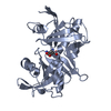 3ekmC 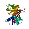 2gkeS S: Starting model for refinement C: citing same article ( |
|---|---|
| Similar structure data |


-Links
 PDBj
PDBj- Assembly
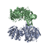
| Deposited unit | 
| ||||||||
|---|---|---|---|---|---|---|---|---|---|
| 1 |
| ||||||||
| 2 | 
| ||||||||
| 3 | 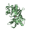
| ||||||||
| 4 | 
| ||||||||
| 5 | 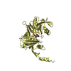
| ||||||||
| 6 | 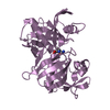
| ||||||||
| 7 | 
| ||||||||
| Unit cell |
|
-Components
| #1: Protein | Mass: 34306.828 Da / Num. of mol.: 6 / Fragment: UNP residues 52-362 Source method: isolated from a genetically manipulated source Source: (gene. exp.)  #2: Chemical | ChemComp-ZDP / (   Mass: 204.224 Da / Num. of mol.: 6 / Source method: obtained synthetically / Formula: C8H16N2O4 Mass: 204.224 Da / Num. of mol.: 6 / Source method: obtained synthetically / Formula: C8H16N2O4#3: Water | ChemComp-HOH / |  Mass: 18.015 Da / Num. of mol.: 1799 / Source method: isolated from a natural source / Formula: H2O Mass: 18.015 Da / Num. of mol.: 1799 / Source method: isolated from a natural source / Formula: H2O |
|---|
-Experimental details
-Experiment
| Experiment | Method: X-RAY DIFFRACTION / Number of used crystals: 1 |
|---|
- Sample preparation
| Crystal | Density Matthews: 3.03 Å3/Da / Density % sol: 59.44 % |
|---|---|
| Crystal grow | Temperature: 300 K / Method: vapor diffusion, hanging drop / pH: 7 Details: 60% v/v Tacsimate buffer, pH 7.0, VAPOR DIFFUSION, HANGING DROP, temperature 300K |
-Data collection
| Diffraction | Mean temperature: 100 K |
|---|---|
| Diffraction source | Source: SYNCHROTRON / Site: ALS  / Beamline: 8.3.1 / Wavelength: 1.115879 Å / Beamline: 8.3.1 / Wavelength: 1.115879 Å |
| Detector | Type: ADSC QUANTUM 210 / Detector: CCD / Date: Mar 13, 2006 / Details: double crystal monochromator |
| Radiation | Monochromator: double crystal / Protocol: SINGLE WAVELENGTH / Monochromatic (M) / Laue (L): M / Scattering type: x-ray |
| Radiation wavelength | Wavelength: 1.115879 Å / Relative weight: 1 |
| Reflection | Resolution: 1.95→50 Å / Num. all: 175374 / Num. obs: 175374 / % possible obs: 98 % / Observed criterion σ(F): 0 / Observed criterion σ(I): 0 / Rmerge(I) obs: 0.068 / Net I/σ(I): 12.2 |
| Reflection shell | Resolution: 1.95→2.02 Å / Rmerge(I) obs: 0.307 / Mean I/σ(I) obs: 2.6 / % possible all: 97.3 |
- Processing
| Software |
| ||||||||||||||||||||
|---|---|---|---|---|---|---|---|---|---|---|---|---|---|---|---|---|---|---|---|---|---|
| Refinement | Method to determine structure: MOLECULAR REPLACEMENT Starting model: 2GKE Resolution: 1.95→50 Å / σ(F): 0 / σ(I): 0 / Stereochemistry target values: Engh & Huber
| ||||||||||||||||||||
| Refine analyze | Luzzati coordinate error obs: 0.2 Å | ||||||||||||||||||||
| Refinement step | Cycle: LAST / Resolution: 1.95→50 Å
| ||||||||||||||||||||
| Refine LS restraints |
|
