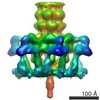
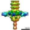
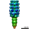
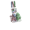
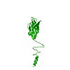
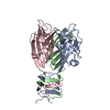
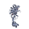
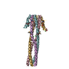
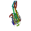
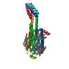
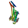
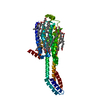
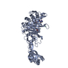
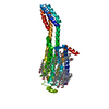
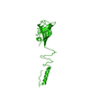
ジャーナル: J Biol Chem / 年: 2010 タイトル: Structure and molecular assignment of lactococcal phage TP901-1 baseplate. 著者: Cecilia Bebeacua / Patrick Bron / Livia Lai / Christina Skovgaard Vegge / Lone Brøndsted / Silvia Spinelli / Valérie Campanacci / David Veesler / Marin van Heel / Christian Cambillau / 要旨: P335 lactococcal phages infect the gram(+) bacterium Lactococcus lactis using a large multiprotein complex located at the distal part of the tail and termed baseplate (BP). The BP harbors the ...P335 lactococcal phages infect the gram(+) bacterium Lactococcus lactis using a large multiprotein complex located at the distal part of the tail and termed baseplate (BP). The BP harbors the receptor-binding proteins (RBPs), which allow the specific recognition of saccharidic receptors localized on the host cell surface. We report here the electron microscopic structure of the phage TP901-1 wild-type BP as well as those of two mutants bppL (-) and bppU(-), lacking BppL (the RBPs) or both peripheral BP components (BppL and BppU), respectively. We also achieved an electron microscopic reconstruction of a partial BP complex, formed by BppU and BppL. This complex exhibits a tripod shape and is composed of nine BppLs and three BppUs. These structures, combined with light-scattering measurements, led us to propose that the TP901-1 BP harbors six tripods at its periphery, located around the central tube formed by ORF46 (Dit) hexamers, at its proximal end, and a ORF47 (Tal) trimer at its distal extremity. A total of 54 BppLs (18 RBPs) are thus available to mediate host anchoring with a large apparent avidity. TP901-1 BP exhibits an infection-ready conformation and differs strikingly from the lactococcal phage p2 BP, bearing only 6 RBPs, and which needs a conformational change to reach its activated state. The comparison of several Siphoviridae structures uncovers a close organization of their central BP core whereas striking differences occur at the periphery, leading to diverse mechanisms of host recognition.
温度: 293 K / 手法: 蒸気拡散法, シッティングドロップ法 / pH: 6.5 詳細: 300 nL of protein at 5 mg/mL were mixed with 100 nL of 20% PEG 8000, 0.2 M Mg Acetate tetrahydrate, 0.1 M Na Cacodylate pH 6.5 using a Cartesian Pixsys Robot, VAPOR DIFFUSION, SITTING DROP, temperature 293K
 ムービー
ムービー コントローラー


 データを開く
 基本情報
基本情報 要素
要素 キーワード
キーワード 機能・相同性情報
 Lactococcus phage TP901-1 (ファージ)
 X線回折 /
  データ登録者
 引用
引用
 構造の表示
構造の表示 ダウンロードとリンク
 その他のダウンロード
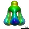
 PDBj
PDBj
 集合体
集合体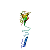

 分子量: 18.015 Da / 分子数: 138 / 由来タイプ: 天然 / 式: H2O
 試料調製
試料調製 / ビームライン: ID14-2 / 波長: 0.933 Å
 解析
解析