 Movie
Movie Controller

[English] 日本語
 Yorodumi
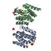
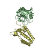
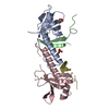
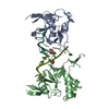
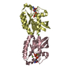
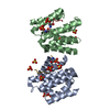
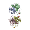
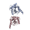
- PDB-3egv: Ribosomal protein L11 methyltransferase (PrmA) in complex with tr... -
+ Open data

- Basic information
| Entry | Database: PDB / ID: 3egv | ||||||||||||
|---|---|---|---|---|---|---|---|---|---|---|---|---|---|
| Title | Ribosomal protein L11 methyltransferase (PrmA) in complex with trimethylated ribosomal protein L11 | ||||||||||||
 Components Components |
| ||||||||||||
 Keywords Keywords | TRANSFERASE/RIBOSOMAL PROTEIN / post-translational modification / multiple methyltransferase / Methyltransferase / Transferase / Methylation / Ribonucleoprotein / Ribosomal protein / RNA-binding / rRNA-binding / TRANSFERASE-RIBOSOMAL PROTEIN COMPLEX | ||||||||||||
| Function / homology |  Function and homology information Function and homology informationprotein-lysine N-methyltransferase activity / Transferases; Transferring one-carbon groups; Methyltransferases / large ribosomal subunit rRNA binding / methylation / cytosolic large ribosomal subunit / structural constituent of ribosome / translation / cytoplasm Similarity search - Function | ||||||||||||
| Biological species |   Thermus thermophilus HB8 (bacteria) Thermus thermophilus HB8 (bacteria) | ||||||||||||
| Method |  X-RAY DIFFRACTION / SYNCHROTRON / MOLECULAR REPLACEMENT / Resolution: 1.75 Å X-RAY DIFFRACTION / SYNCHROTRON / MOLECULAR REPLACEMENT / Resolution: 1.75 Å | ||||||||||||
 Authors Authors | Demirci, H. / Gregory, S.T. / Dahlberg, A.E. / Jogl, G. | ||||||||||||
 Citation Citation | Journal: Structure / Year: 2008 Title: Multiple-site trimethylation of ribosomal protein L11 by the PrmA methyltransferase. Authors: Demirci, H. / Gregory, S.T. / Dahlberg, A.E. / Jogl, G. | ||||||||||||
| History |
|
- Structure visualization
| Structure viewer | Molecule: MolmilJmol/JSmol |
|---|
- Downloads & links
-Download
| PDBx/mmCIF format | 3egv.cif.gz | 92.9 KB | Display | PDBx/mmCIF format |
|---|---|---|---|---|
| PDB format | pdb3egv.ent.gz | 67.8 KB | Display | PDB format |
| PDBx/mmJSON format | 3egv.json.gz | Tree view | PDBx/mmJSON format | |
| Others |  Other downloads Other downloads |
-Validation report
| Arichive directory | https://data.pdbj.org/pub/pdb/validation_reports/eg/3egvftp://data.pdbj.org/pub/pdb/validation_reports/eg/3egv | HTTPS FTP |
|---|
-Related structure data
| Related structure data | 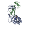 3cjqC 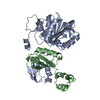 3cjrC  3cjsC 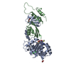 3cjtC  2nxcS  2nxnS S: Starting model for refinement C: citing same article ( |
|---|---|
| Similar structure data |


-Links
 PDBj
PDBj


- Assembly
| Deposited unit | 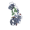
| ||||||||
|---|---|---|---|---|---|---|---|---|---|
| 1 |
| ||||||||
| Unit cell |
|
-Components
-Protein , 2 types, 2 molecules AB
| #1: Protein | Mass: 27661.807 Da / Num. of mol.: 1 Source method: isolated from a genetically manipulated source Details: cytoplasm / Source: (gene. exp.) Thermus thermophilus HB8 (bacteria) / Strain: HB8 / Gene: prmA, TTHA0656 / Plasmid: pET30b / Production host: References: UniProt: Q84BQ9, Transferases; Transferring one-carbon groups; Methyltransferases |
|---|---|
| #2: Protein | Mass: 15511.098 Da / Num. of mol.: 1 / Mutation: K16A Source method: isolated from a genetically manipulated source Details: cytoplasm / Source: (gene. exp.) Thermus thermophilus HB8 (bacteria) / Strain: HB8 / Gene: rpl11, rplK, TTHA0247 / Plasmid: pET11a / Production host: |
-Non-polymers , 5 types, 422 molecules 


| #3: Chemical | ChemComp-SAH /  Type: L-peptide linking / Mass: 384.411 Da / Num. of mol.: 1 / Source method: obtained synthetically / Formula: C14H20N6O5S Type: L-peptide linking / Mass: 384.411 Da / Num. of mol.: 1 / Source method: obtained synthetically / Formula: C14H20N6O5S | ||||||
|---|---|---|---|---|---|---|---|
| #4: Chemical | ChemComp-GOL /  Mass: 92.094 Da / Num. of mol.: 4 / Source method: obtained synthetically / Formula: C3H8O3 Mass: 92.094 Da / Num. of mol.: 4 / Source method: obtained synthetically / Formula: C3H8O3#5: Chemical | ChemComp-CL / |  Mass: 35.453 Da / Num. of mol.: 1 / Source method: obtained synthetically / Formula: Cl Mass: 35.453 Da / Num. of mol.: 1 / Source method: obtained synthetically / Formula: Cl#6: Chemical | ChemComp-IOD / |  Mass: 126.904 Da / Num. of mol.: 1 / Source method: obtained synthetically / Formula: I Mass: 126.904 Da / Num. of mol.: 1 / Source method: obtained synthetically / Formula: I#7: Water | ChemComp-HOH / | Mass: 18.015 Da / Num. of mol.: 415 / Source method: isolated from a natural source / Formula: H2O |
-Experimental details
-Experiment
| Experiment | Method: X-RAY DIFFRACTION / Number of used crystals: 1 |
|---|
- Sample preparation
| Crystal | Density Matthews: 2.97 Å3/Da / Density % sol: 58.64 % |
|---|---|
| Crystal grow | Temperature: 277 K / Method: microbatch technique under oil / pH: 6.5 Details: 160 mM calcium acetate hydrate, 80 mM sodium cacodylate, 14.4% w/v PEG8000, 20% v/v glycerol, 4mM AdoMet, pH 6.5, microbatch technique under oil, temperature 277.0K |
-Data collection
| Diffraction | Mean temperature: 100 K |
|---|---|
| Diffraction source | Source: SYNCHROTRON / Site: NSLS  / Beamline: X4A / Wavelength: 0.9797 Å / Beamline: X4A / Wavelength: 0.9797 Å |
| Detector | Type: ADSC QUANTUM 4 / Detector: CCD / Date: Sep 15, 2006 Details: Variable vertical and fixed horizontal slits. KOHZU double crystal monochromator with a water-cooled flat first crystal and a sagittally focused second crystal positioned for a fixed exit ...Details: Variable vertical and fixed horizontal slits. KOHZU double crystal monochromator with a water-cooled flat first crystal and a sagittally focused second crystal positioned for a fixed exit beam condition. Located ~18 m from source and ~6 m from sample position. Mirror system consisting of two vertically stacked, fused silica, spherical mirrors, to provide vertical focusing and harmonic rejection. One of the mirrors is rhodium coated and the other is uncoated. Located ~19.7 m from source. |
| Radiation | Monochromator: KOHZU double crystal monochromator with a water-cooled flat first crystal and a sagittally focused second crystal positioned for a fixed exit beam condition. Located ~18 m from source ...Monochromator: KOHZU double crystal monochromator with a water-cooled flat first crystal and a sagittally focused second crystal positioned for a fixed exit beam condition. Located ~18 m from source and ~6 m from sample position. Protocol: SINGLE WAVELENGTH / Monochromatic (M) / Laue (L): M / Scattering type: x-ray |
| Radiation wavelength | Wavelength: 0.9797 Å / Relative weight: 1 |
| Reflection | Resolution: 1.75→30 Å / Num. obs: 48051 / % possible obs: 97.2 % / Observed criterion σ(I): -3 / Redundancy: 1.7 % / Rmerge(I) obs: 0.056 / Net I/σ(I): 12.2 |
| Reflection shell | Resolution: 1.75→1.81 Å / Redundancy: 1.6 % / Rmerge(I) obs: 0.334 / Mean I/σ(I) obs: 2 / Num. unique all: 9414 / % possible all: 93.8 |
- Processing
| Software |
| ||||||||||||||||||||||||||||||||||||||||||||||||||||||||||||||||||||||||||||||||||||||||||||||||||||||||||||||||||||||||||||||||||||||||||||||||||||||||||||||||||||||||||
|---|---|---|---|---|---|---|---|---|---|---|---|---|---|---|---|---|---|---|---|---|---|---|---|---|---|---|---|---|---|---|---|---|---|---|---|---|---|---|---|---|---|---|---|---|---|---|---|---|---|---|---|---|---|---|---|---|---|---|---|---|---|---|---|---|---|---|---|---|---|---|---|---|---|---|---|---|---|---|---|---|---|---|---|---|---|---|---|---|---|---|---|---|---|---|---|---|---|---|---|---|---|---|---|---|---|---|---|---|---|---|---|---|---|---|---|---|---|---|---|---|---|---|---|---|---|---|---|---|---|---|---|---|---|---|---|---|---|---|---|---|---|---|---|---|---|---|---|---|---|---|---|---|---|---|---|---|---|---|---|---|---|---|---|---|---|---|---|---|---|---|---|
| Refinement | Method to determine structure: MOLECULAR REPLACEMENT Starting model: pdb entries 2NXC, 2NXN Resolution: 1.75→30 Å / Cor.coef. Fo:Fc: 0.961 / Cor.coef. Fo:Fc free: 0.949 / SU B: 3.55 / SU ML: 0.063 / TLS residual ADP flag: LIKELY RESIDUAL / Cross valid method: THROUGHOUT / ESU R: 0.094 / ESU R Free: 0.095 / Stereochemistry target values: MAXIMUM LIKELIHOOD / Details: HYDROGENS HAVE BEEN ADDED IN THE RIDING POSITIONS
| ||||||||||||||||||||||||||||||||||||||||||||||||||||||||||||||||||||||||||||||||||||||||||||||||||||||||||||||||||||||||||||||||||||||||||||||||||||||||||||||||||||||||||
| Solvent computation | Ion probe radii: 0.8 Å / Shrinkage radii: 0.8 Å / VDW probe radii: 1.4 Å / Solvent model: MASK | ||||||||||||||||||||||||||||||||||||||||||||||||||||||||||||||||||||||||||||||||||||||||||||||||||||||||||||||||||||||||||||||||||||||||||||||||||||||||||||||||||||||||||
| Displacement parameters | Biso mean: 33.628 Å2
| ||||||||||||||||||||||||||||||||||||||||||||||||||||||||||||||||||||||||||||||||||||||||||||||||||||||||||||||||||||||||||||||||||||||||||||||||||||||||||||||||||||||||||
| Refinement step | Cycle: LAST / Resolution: 1.75→30 Å
| ||||||||||||||||||||||||||||||||||||||||||||||||||||||||||||||||||||||||||||||||||||||||||||||||||||||||||||||||||||||||||||||||||||||||||||||||||||||||||||||||||||||||||
| Refine LS restraints |
| ||||||||||||||||||||||||||||||||||||||||||||||||||||||||||||||||||||||||||||||||||||||||||||||||||||||||||||||||||||||||||||||||||||||||||||||||||||||||||||||||||||||||||
| LS refinement shell | Resolution: 1.75→1.794 Å / Total num. of bins used: 20
| ||||||||||||||||||||||||||||||||||||||||||||||||||||||||||||||||||||||||||||||||||||||||||||||||||||||||||||||||||||||||||||||||||||||||||||||||||||||||||||||||||||||||||
| Refinement TLS params. | Method: refined / Refine-ID: X-RAY DIFFRACTION
| ||||||||||||||||||||||||||||||||||||||||||||||||||||||||||||||||||||||||||||||||||||||||||||||||||||||||||||||||||||||||||||||||||||||||||||||||||||||||||||||||||||||||||
| Refinement TLS group |
|
