 Movie
Movie Controller

[English] 日本語
 Yorodumi
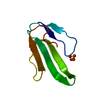
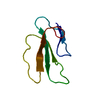
- PDB-3ebx: REFINEMENT AT 1.4 ANGSTROMS RESOLUTION OF A MODEL OF ERABUTOXIN B... -
+ Open data

- Basic information
| Entry | Database: PDB / ID: 3ebx | |||||||||
|---|---|---|---|---|---|---|---|---|---|---|
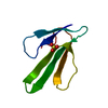
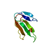
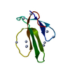
| Title | REFINEMENT AT 1.4 ANGSTROMS RESOLUTION OF A MODEL OF ERABUTOXIN B. TREATMENT OF ORDERED SOLVENT AND DISCRETE DISORDER | |||||||||
 Components Components | ERABUTOXIN B | |||||||||
 Keywords Keywords | TOXIN | |||||||||
| Function / homology |  Function and homology information Function and homology informationacetylcholine receptor inhibitor activity / ion channel regulator activity / toxin activity / extracellular region Similarity search - Function | |||||||||
| Biological species |  Laticauda semifasciata (broad-banded blue sea krait) Laticauda semifasciata (broad-banded blue sea krait) | |||||||||
| Method |  X-RAY DIFFRACTION / Resolution: 1.4 Å X-RAY DIFFRACTION / Resolution: 1.4 Å | |||||||||
 Authors Authors | Smith, J.L. / Corfield, P.W.R. / Hendrickson, W.A. / Low, B.W. | |||||||||
 Citation Citation | Journal: Acta Crystallogr.,Sect.A / Year: 1988 Title: Refinement at 1.4 A resolution of a model of erabutoxin b: treatment of ordered solvent and discrete disorder. Authors: Smith, J.L. / Corfield, P.W. / Hendrickson, W.A. / Low, B.W. #1: Journal: Asia Pac.J.Pharmacol. / Year: 1987Title: Acetylcholine Receptor. Alpha-Toxin Binding Site-Theoretical and Model Studies Authors: Low, B.W. / Corfield, P.W.R. #2: Journal: Eur.J.Biochem. / Year: 1986Title: Erabutoxin B. Structure(Slash)Function Relationships Following Initial Protein Refinement at 0.140-Nm Resolution Authors: Low, B.W. / Corfield, P.W.R. #3: Journal: Eur.J.Biochem. / Year: 1985Title: Erabutoxin B. Initial Protein Refinement and Sequence Analysis at 0.140-Nm Resolution Authors: Bourne, P.E. / Sato, A. / Corfield, P.W.R. / Rosen, L.S. / Birken, S. / Low, B.W. #4: Journal: J.Biol.Chem. / Year: 1980Title: The Toxin-Agglutinin Fold. A New Group of Small Protein Structures Organized Around a Four-Disulfide Core Authors: Drenth, J. / Low, B.W. / Richardson, J.S. / Wright, C.S. #5: Journal: Biochem.Biophys.Res.Commun. / Year: 1979Title: Molecular Conformation of Erabutoxin B. Atomic Coordinates at 2.5 Angstroms Resolution Authors: Kimball, M.R. / Sato, A. / Richardson, J.S. / Rosen, L.S. / Low, B.W. #6: Journal: Handb.Exp.Pharmacol. / Year: 1979Title: The Three-Dimensional Structure of Postsynaptic Snake Neurotoxins. Consideration of Structure and Function Authors: Low, B.W. #7: Journal: Proc.Natl.Acad.Sci.USA / Year: 1976Title: Three Dimensional Structure of Erabutoxin B Neurotoxic Protein. Inhibitor of Acetylcholine Receptor Authors: Low, B.W. / Preston, H.S. / Sato, A. / Rosen, L.S. / Searl, J.E. / Rudko, A.D. / Richardson, J.S. #8: Journal: J.Biol.Chem. / Year: 1971Title: X-Ray Crystallographic Study of the Erabutoxins and of a Diiodo Derivative Authors: Low, B.W. / Potter, R. / Jackson, R.B. / Tamiya, N. / Sato, S. | |||||||||
| History |
| |||||||||
| Remark 700 | SHEET THE BETA SHEET *DCE* WAS INADVERTENTLY NOT INCLUDED IN ENTRY *2EBX*. |
- Structure visualization
| Structure viewer | Molecule: MolmilJmol/JSmol |
|---|
- Downloads & links
-Download
| PDBx/mmCIF format | 3ebx.cif.gz | 30 KB | Display | PDBx/mmCIF format |
|---|---|---|---|---|
| PDB format | pdb3ebx.ent.gz | 19.8 KB | Display | PDB format |
| PDBx/mmJSON format | 3ebx.json.gz | Tree view | PDBx/mmJSON format | |
| Others |  Other downloads Other downloads |
-Validation report
| Arichive directory | https://data.pdbj.org/pub/pdb/validation_reports/eb/3ebxftp://data.pdbj.org/pub/pdb/validation_reports/eb/3ebx | HTTPS FTP |
|---|
-Related structure data
| Related structure data | |
|---|---|
| Similar structure data |


-Links
 PDBj
PDBj

- Assembly
| Deposited unit | 
| ||||||||
|---|---|---|---|---|---|---|---|---|---|
| 1 |
| ||||||||
| Unit cell |
| ||||||||
| Atom site foot note | 1: SEE REMARK 4. |
-Components
| #1: Protein | Mass: 6877.759 Da / Num. of mol.: 1 Source method: isolated from a genetically manipulated source Source: (gene. exp.) Laticauda semifasciata (broad-banded blue sea krait)References: UniProt: Q90VW1 |
|---|---|
| #2: Chemical | ChemComp-SO4 /   Mass: 96.063 Da / Num. of mol.: 1 / Source method: obtained synthetically / Formula: SO4 Mass: 96.063 Da / Num. of mol.: 1 / Source method: obtained synthetically / Formula: SO4 |
| #3: Water | ChemComp-HOH /  Mass: 18.015 Da / Num. of mol.: 111 / Source method: isolated from a natural source / Formula: H2O Mass: 18.015 Da / Num. of mol.: 111 / Source method: isolated from a natural source / Formula: H2O |
| Has protein modification | Y |
| Sequence details | THE POSTSYNAPTIC NEUROTOXINS OF SEA SNAKE VENOM ARE ANTAGONISTS OF THE NICOTINIC ACETYLCHOLINE ...THE POSTSYNAPT |
| Source details | THE PREVIOUS REFINEMENT ESTABLISHED THE STRUCTURAL IDENTITY OF ERABUTOXIN B AND NEUROTOXIN B. ...THE PREVIOUS REFINEMENT |
-Experimental details
-Experiment
| Experiment | Method: X-RAY DIFFRACTION |
|---|
- Sample preparation
| Crystal | Density Matthews: 1.82 Å3/Da / Density % sol: 32.58 % | |||||||||||||||
|---|---|---|---|---|---|---|---|---|---|---|---|---|---|---|---|---|
| Crystal grow | *PLUS pH: 7.5 / Method: unknown / Details: referred to FEBS Lett.68.1-4 1976 | |||||||||||||||
| Components of the solutions | *PLUS
|
-Data collection
| Radiation | Scattering type: x-ray |
|---|---|
| Radiation wavelength | Relative weight: 1 |
| Reflection | *PLUS |
- Processing
| Software | Name: PROLSQ / Classification: refinement | ||||||||||||||||||||||||||||||||||||||||||||
|---|---|---|---|---|---|---|---|---|---|---|---|---|---|---|---|---|---|---|---|---|---|---|---|---|---|---|---|---|---|---|---|---|---|---|---|---|---|---|---|---|---|---|---|---|---|
| Refinement | Resolution: 1.4→10 Å / σ(F): 2 Details: SHIFTS FROM ENTRY *2EBX* IN POSITIONS OF NON-DISORDERED PROTEIN ATOMS ARE VERY SMALL. THE RMS DEVIATION IS 0.5 ANGSTROMS FOR ALL NON-DISORDERED ATOMS AND 0.07 ANGSTROMS FOR MAIN CHAIN ATOMS.
| ||||||||||||||||||||||||||||||||||||||||||||
| Refinement step | Cycle: LAST / Resolution: 1.4→10 Å
| ||||||||||||||||||||||||||||||||||||||||||||
| Refine LS restraints |
| ||||||||||||||||||||||||||||||||||||||||||||
| Refinement | *PLUS Highest resolution: 1.4 Å / Lowest resolution: 10 Å / Num. reflection obs: 7732 / σ(F): 2 / Rfactor obs: 0.14 | ||||||||||||||||||||||||||||||||||||||||||||
| Solvent computation | *PLUS | ||||||||||||||||||||||||||||||||||||||||||||
| Displacement parameters | *PLUS | ||||||||||||||||||||||||||||||||||||||||||||
| Refine LS restraints | *PLUS
|
