 Movie
Movie Controller


+ Open data

- Basic information
| Entry | Database: PDB / ID: 2era | ||||||
|---|---|---|---|---|---|---|---|
| Title | RECOMBINANT ERABUTOXIN A, S8G MUTANT | ||||||
 Components Components | ERABUTOXIN A | ||||||
 Keywords Keywords | NEUROTOXIN / SNAKE NEUROTOXIN / VENOM / POSTSYNAPTIC NEUROTOXIN | ||||||
| Function / homology |  Function and homology information Function and homology informationacetylcholine receptor inhibitor activity / ion channel regulator activity / toxin activity / extracellular region Similarity search - Function | ||||||
| Biological species |  Laticauda semifasciata (broad-banded blue sea krait) Laticauda semifasciata (broad-banded blue sea krait) | ||||||
| Method |  X-RAY DIFFRACTION / SYNCHROTRON / MOLECULAR REPLACEMENT / Resolution: 1.81 Å X-RAY DIFFRACTION / SYNCHROTRON / MOLECULAR REPLACEMENT / Resolution: 1.81 Å | ||||||
 Authors Authors | Gaucher, J.F. / Menez, R. / Arnoux, B. / Menez, A. / Ducruix, A. | ||||||
 Citation Citation | Journal: Eur.J.Biochem. / Year: 2000 Title: High resolution x-ray analysis of two mutants of a curaremimetic snake toxin Authors: Gaucher, J.F. / Menez, R. / Arnoux, B. / Menez, A. / Ducruix, A. #1: Journal: J.Biol.Chem. / Year: 1995Title: Genetic Engineering of Snake Toxins. The Functional Site of Erabutoxin A, as Delineated by Site-Directed Mutagenesis, Includes Variant Residues Authors: Tremeau, O. / Lemaire, C. / Drevet, P. / Pinkasfeld, S. / Ducancel, F. / Boulain, J.C. / Menez, A. #2: Journal: FEBS Lett. / Year: 1994Title: Three-Dimensional Crystal Structure of Recombinant Erabutoxin a at 2.0 A Resolution Authors: Arnoux, B. / Menez, R. / Drevet, P. / Boulain, J.C. / Ducruix, A. / Menez, A. #3: Journal: J.Biol.Chem. / Year: 1993Title: Genetic Engineering of Snake Toxins. Role of Invariant Residues in the Structural and Functional Properties of a Curaremimetic Toxin, as Probed by Site-Directed Mutagenesis Authors: Pillet, L. / Tremeau, O. / Ducancel, F. / Drevet, P. / Zinn-Justin, S. / Pinkasfeld, S. / Boulain, J.C. / Menez, A. #4: Journal: J.Biol.Chem. / Year: 1989Title: The Crystal Structure of Erabutoxin a at 2.0-A Resolution Authors: Corfield, P.W. / Lee, T.J. / Low, B.W. #5: Journal: Acta Crystallogr.,Sect.A / Year: 1988Title: Refinement at 1.4 A Resolution of a Model of Erabutoxin B: Treatment of Ordered Solvent and Discrete Disorder Authors: Smith, J.L. / Corfield, P.W.R. / Hendrickson, W.A. / Low, B.W. | ||||||
| History |
|
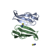
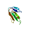
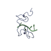
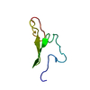
- Structure visualization
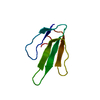
| Structure viewer | Molecule: MolmilJmol/JSmol |
|---|
- Downloads & links
-Download
| PDBx/mmCIF format | 2era.cif.gz | 25.6 KB | Display | PDBx/mmCIF format |
|---|---|---|---|---|
| PDB format | pdb2era.ent.gz | 15.5 KB | Display | PDB format |
| PDBx/mmJSON format | 2era.json.gz | Tree view | PDBx/mmJSON format | |
| Others |  Other downloads Other downloads |
-Validation report
| Arichive directory | https://data.pdbj.org/pub/pdb/validation_reports/er/2eraftp://data.pdbj.org/pub/pdb/validation_reports/er/2era | HTTPS FTP |
|---|
-Related structure data


-Links
 PDBj
PDBj

- Assembly
| Deposited unit | 
| ||||||||
|---|---|---|---|---|---|---|---|---|---|
| 1 |
| ||||||||
| Unit cell |
|
-Components
| #1: Protein | Mass: 6823.689 Da / Num. of mol.: 1 / Mutation: S8G Source method: isolated from a genetically manipulated source Source: (gene. exp.) Laticauda semifasciata (broad-banded blue sea krait)Variant: TYPE A (N26 AND K51) / Plasmid: PRIT5 / Production host:  |
|---|---|
| #2: Water | ChemComp-HOH /  Mass: 18.015 Da / Num. of mol.: 61 / Source method: isolated from a natural source / Formula: H2O Mass: 18.015 Da / Num. of mol.: 61 / Source method: isolated from a natural source / Formula: H2O |
| Has protein modification | Y |
-Experimental details
-Experiment
| Experiment | Method: X-RAY DIFFRACTION / Number of used crystals: 1 |
|---|
- Sample preparation
| Crystal | Density Matthews: 1.8 Å3/Da / Density % sol: 35 % | ||||||||||||||||||||||||
|---|---|---|---|---|---|---|---|---|---|---|---|---|---|---|---|---|---|---|---|---|---|---|---|---|---|
| Crystal grow | Method: vapor diffusion, hanging drop / pH: 4.5 Details: CRYSTALLIZATION WERE PERFORMED AT 291K BY THE HANGING DROP METHOD. DROPS OF 2 MICROLITRE OF 0.007M PROTEIN AND 2 MICROLITRE OF RESERVOIR WERE EQUILIBRATED AGAINST 3M NACL, 0.05M NAOAC BUFFER ...Details: CRYSTALLIZATION WERE PERFORMED AT 291K BY THE HANGING DROP METHOD. DROPS OF 2 MICROLITRE OF 0.007M PROTEIN AND 2 MICROLITRE OF RESERVOIR WERE EQUILIBRATED AGAINST 3M NACL, 0.05M NAOAC BUFFER SOLUTION (PH 4.5)., vapor diffusion - hanging drop | ||||||||||||||||||||||||
| Crystal grow | *PLUS Temperature: 18 ℃ / Method: vapor diffusionDetails: drop solution was mixed with an equal volume of reservoir solution | ||||||||||||||||||||||||
| Components of the solutions | *PLUS
|
-Data collection
| Diffraction | Mean temperature: 278 K |
|---|---|
| Diffraction source | Source: SYNCHROTRON / Site: LURE  / Beamline: DW32 / Wavelength: 0.901 / Beamline: DW32 / Wavelength: 0.901 |
| Detector | Type: MARRESEARCH / Detector: IMAGE PLATE / Date: Sep 1, 1993 / Details: MIRRORS |
| Radiation | Monochromatic (M) / Laue (L): M / Scattering type: x-ray |
| Radiation wavelength | Wavelength: 0.901 Å / Relative weight: 1 |
| Reflection | Resolution: 1.81→15 Å / Num. obs: 4889 / % possible obs: 99.8 % / Observed criterion σ(I): 3 / Redundancy: 8.4 % / Biso Wilson estimate: 10.8 Å2 / Rsym value: 0.047 / Net I/σ(I): 13 |
| Reflection shell | Resolution: 1.814→1.86 Å / Redundancy: 5.8 % / Mean I/σ(I) obs: 6.3 / Rsym value: 0.116 / % possible all: 97.6 |
| Reflection | *PLUS Lowest resolution: 17 Å / Num. obs: 4924 / % possible obs: 99 % / Num. measured all: 41373 / Rmerge(I) obs: 0.047 |
| Reflection shell | *PLUS % possible obs: 97 % / Rmerge(I) obs: 0.116 |
- Processing
| Software |
| ||||||||||||||||||||||||||||||||||||||||||||||||||||||||||||
|---|---|---|---|---|---|---|---|---|---|---|---|---|---|---|---|---|---|---|---|---|---|---|---|---|---|---|---|---|---|---|---|---|---|---|---|---|---|---|---|---|---|---|---|---|---|---|---|---|---|---|---|---|---|---|---|---|---|---|---|---|---|
| Refinement | Method to determine structure: MOLECULAR REPLACEMENT Starting model: STRUCTURE OF RECOMBINANT ERABUTOXIN A PROVIDED BY DR.B.ARNOUX Resolution: 1.81→10 Å / Rfactor Rfree error: 0.01 / Data cutoff high absF: 100000 / Data cutoff low absF: 0 / Isotropic thermal model: RESTRAINED / Cross valid method: THROUGHOUT / σ(F): 2 Details: AT THE END OF REFINEMENT THE WHOLE SET OF DATA WAS USED FOR THE LAST STEPS OF REFINEMENT.
| ||||||||||||||||||||||||||||||||||||||||||||||||||||||||||||
| Displacement parameters | Biso mean: 12.1 Å2 | ||||||||||||||||||||||||||||||||||||||||||||||||||||||||||||
| Refine analyze | Luzzati coordinate error obs: 0.18 Å / Luzzati d res low obs: 10 Å / Luzzati sigma a obs: 0.08 Å | ||||||||||||||||||||||||||||||||||||||||||||||||||||||||||||
| Refinement step | Cycle: LAST / Resolution: 1.81→10 Å
| ||||||||||||||||||||||||||||||||||||||||||||||||||||||||||||
| Refine LS restraints |
| ||||||||||||||||||||||||||||||||||||||||||||||||||||||||||||
| LS refinement shell | Resolution: 1.81→1.88 Å / Rfactor Rfree error: 0.032 / Total num. of bins used: 8
| ||||||||||||||||||||||||||||||||||||||||||||||||||||||||||||
| Xplor file |
| ||||||||||||||||||||||||||||||||||||||||||||||||||||||||||||
| Software | *PLUS Name: X-PLOR / Version: 3.1 / Classification: refinement | ||||||||||||||||||||||||||||||||||||||||||||||||||||||||||||
| Refinement | *PLUS | ||||||||||||||||||||||||||||||||||||||||||||||||||||||||||||
| Solvent computation | *PLUS | ||||||||||||||||||||||||||||||||||||||||||||||||||||||||||||
| Displacement parameters | *PLUS | ||||||||||||||||||||||||||||||||||||||||||||||||||||||||||||
| Refine LS restraints | *PLUS
| ||||||||||||||||||||||||||||||||||||||||||||||||||||||||||||
| LS refinement shell | *PLUS Rfactor obs: 0.193 |
