 Movie
Movie Controller

[English] 日本語
 Yorodumi
- PDB-3e9w: X-Ray Crystal Structure of the hexamer DCACACG:Crystal grown in t... -
+ Open data

- Basic information
| Entry | Database: PDB / ID: 3e9w | ||||||
|---|---|---|---|---|---|---|---|
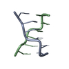
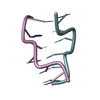
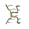
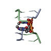
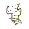
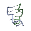
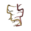
| Title | X-Ray Crystal Structure of the hexamer DCACACG:Crystal grown in the presence of cobalt(III)hexammine Chloride. | ||||||
 Components Components |
| ||||||
 Keywords Keywords | DNA / Z DOUBLE HELIX | ||||||
| Function / homology | DNA Function and homology information Function and homology information | ||||||
| Method |  X-RAY DIFFRACTION / MOLECULAR REPLACEMENT / Resolution: 2.05 Å X-RAY DIFFRACTION / MOLECULAR REPLACEMENT / Resolution: 2.05 Å | ||||||
 Authors Authors | Venkadesh, S. / Mandal, P.K. / Gautham, N. | ||||||
 Citation Citation | Journal: Acta Crystallogr.,Sect.F / Year: 2009 Title: The structure of d(CACACG).d(CGTGTG). Authors: Venkadesh, S. / Mandal, P.K. / Gautham, N. | ||||||
| History |
|
- Structure visualization
| Structure viewer | Molecule: MolmilJmol/JSmol |
|---|
- Downloads & links
-Download
| PDBx/mmCIF format | 3e9w.cif.gz | 18.5 KB | Display | PDBx/mmCIF format |
|---|---|---|---|---|
| PDB format | pdb3e9w.ent.gz | 10.2 KB | Display | PDB format |
| PDBx/mmJSON format | 3e9w.json.gz | Tree view | PDBx/mmJSON format | |
| Others |  Other downloads Other downloads |
-Validation report
| Arichive directory | https://data.pdbj.org/pub/pdb/validation_reports/e9/3e9wftp://data.pdbj.org/pub/pdb/validation_reports/e9/3e9w | HTTPS FTP |
|---|
-Related structure data
| Similar structure data |
|---|


-Links
 PDBj
PDBj

- Assembly
| Deposited unit | 
| ||||||||
|---|---|---|---|---|---|---|---|---|---|
| 1 |
| ||||||||
| Unit cell |
|
-Components
| #1: DNA chain | Mass: 1778.207 Da / Num. of mol.: 1 / Source method: obtained synthetically |
|---|---|
| #2: DNA chain | Mass: 1840.227 Da / Num. of mol.: 1 / Source method: obtained synthetically |
| #3: Water | ChemComp-HOH /  Mass: 18.015 Da / Num. of mol.: 20 / Source method: isolated from a natural source / Formula: H2O Mass: 18.015 Da / Num. of mol.: 20 / Source method: isolated from a natural source / Formula: H2O |
-Experimental details
-Experiment
| Experiment | Method: X-RAY DIFFRACTION / Number of used crystals: 1 |
|---|
- Sample preparation
| Crystal | Density Matthews: 1.67 Å3/Da | ||||||||||||||||||||||||
|---|---|---|---|---|---|---|---|---|---|---|---|---|---|---|---|---|---|---|---|---|---|---|---|---|---|
| Crystal grow | Temperature: 300 K / Method: vapor diffusion, hanging drop / pH: 6.99 Details: cobalt(III)hexammine chloride,spermine and 50% MPD, pH 6.99, VAPOR DIFFUSION, HANGING DROP, temperature 300K | ||||||||||||||||||||||||
| Components of the solutions |
|
-Data collection
| Diffraction | Mean temperature: 300 K |
|---|---|
| Diffraction source | Source: ROTATING ANODE / Type: RIGAKU RU300 / Wavelength: 1.5418 Å |
| Detector | Type: MARRESEARCH / Detector: IMAGE PLATE / Date: Oct 30, 2006 |
| Radiation | Monochromator: MIRRORS / Protocol: SINGLE WAVELENGTH / Monochromatic (M) / Laue (L): M / Scattering type: x-ray |
| Radiation wavelength | Wavelength: 1.5418 Å / Relative weight: 1 |
| Reflection | Resolution: 2.05→15 Å / Num. all: 1384 / Num. obs: 1384 / % possible obs: 92.1 % / Observed criterion σ(F): 0 / Observed criterion σ(I): 0 / Redundancy: 2.73 % / Rmerge(I) obs: 0.027 / Net I/σ(I): 11.7 |
| Reflection shell | Resolution: 2.05→2.13 Å / Redundancy: 2.74 % / Rmerge(I) obs: 0.1984 / Mean I/σ(I) obs: 2.5 / Num. unique all: 120 / % possible all: 94.5 |
- Processing
| Software |
| |||||||||||||||||||||||||||||||||||||||||||||
|---|---|---|---|---|---|---|---|---|---|---|---|---|---|---|---|---|---|---|---|---|---|---|---|---|---|---|---|---|---|---|---|---|---|---|---|---|---|---|---|---|---|---|---|---|---|---|
| Refinement | Method to determine structure: MOLECULAR REPLACEMENT Starting model: Fiber model of Z-DNA Resolution: 2.05→14.58 Å / Cor.coef. Fo:Fc: 0.962 / Cor.coef. Fo:Fc free: 0.952 / SU ML: 0.225 / Isotropic thermal model: Overall / Cross valid method: THROUGHOUT / ESU R: 0.351 / ESU R Free: 0.251 / Stereochemistry target values: MAXIMUM LIKELIHOOD
| |||||||||||||||||||||||||||||||||||||||||||||
| Solvent computation | Ion probe radii: 0.8 Å / Shrinkage radii: 0.8 Å / VDW probe radii: 1.2 Å / Solvent model: MASK | |||||||||||||||||||||||||||||||||||||||||||||
| Displacement parameters | Biso mean: 34.975 Å2
| |||||||||||||||||||||||||||||||||||||||||||||
| Refinement step | Cycle: LAST / Resolution: 2.05→14.58 Å
| |||||||||||||||||||||||||||||||||||||||||||||
| Refine LS restraints |
| |||||||||||||||||||||||||||||||||||||||||||||
| LS refinement shell | Resolution: 2.05→2.107 Å / Total num. of bins used: 20
|
