 Movie
Movie Controller

[English] 日本語
 Yorodumi
- PDB-3e9n: Crystal structure of a putative short-chain dehydrogenase/reducta... -
+ Open data

- Basic information
| Entry | Database: PDB / ID: 3e9n | ||||||
|---|---|---|---|---|---|---|---|
| Title | Crystal structure of a putative short-chain dehydrogenase/reductase from Corynebacterium glutamicum | ||||||
 Components Components | PUTATIVE SHORT-CHAIN DEHYDROGENASE/REDUCTASE | ||||||
 Keywords Keywords | OXIDOREDUCTASE / STRUCTURAL GENOMICS / UNKNOWN FUNCTION / PSI-2 / Protein Structure Initiative / New York SGX Research Center for Structural Genomics / NYSGXRC | ||||||
| Function / homology |  Function and homology information Function and homology informationacetoacetyl-CoA reductase / acetoacetyl-CoA reductase activity / membrane Similarity search - Function | ||||||
| Biological species |  Corynebacterium glutamicum (bacteria) Corynebacterium glutamicum (bacteria) | ||||||
| Method |  X-RAY DIFFRACTION / SYNCHROTRON / SAD / Resolution: 2.4 Å X-RAY DIFFRACTION / SYNCHROTRON / SAD / Resolution: 2.4 Å | ||||||
 Authors Authors | Bonanno, J.B. / Gilmore, M. / Bain, K.T. / Hu, S. / Romero, R. / Smith, D. / Wasserman, S. / Sauder, J.M. / Burley, S.K. / Almo, S.C. / New York SGX Research Center for Structural Genomics (NYSGXRC) | ||||||
 Citation Citation | Journal: To be Published Title: Crystal structure of a putative short-chain dehydrogenase/reductase from Corynebacterium glutamicum Authors: Bonanno, J.B. / Gilmore, M. / Bain, K.T. / Hu, S. / Romero, R. / Smith, D. / Wasserman, S. / Sauder, J.M. / Burley, S.K. / Almo, S.C. | ||||||
| History |
|
- Structure visualization
| Structure viewer | Molecule: MolmilJmol/JSmol |
|---|
- Downloads & links
-Download
| PDBx/mmCIF format | 3e9n.cif.gz | 277 KB | Display | PDBx/mmCIF format |
|---|---|---|---|---|
| PDB format | pdb3e9n.ent.gz | 222.4 KB | Display | PDB format |
| PDBx/mmJSON format | 3e9n.json.gz | Tree view | PDBx/mmJSON format | |
| Others |  Other downloads Other downloads |
-Validation report
| Arichive directory | https://data.pdbj.org/pub/pdb/validation_reports/e9/3e9nftp://data.pdbj.org/pub/pdb/validation_reports/e9/3e9n | HTTPS FTP |
|---|
-Related structure data
| Similar structure data | |
|---|---|
| Other databases |


-Links
 PDBj
PDBj


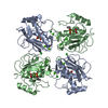
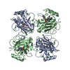
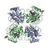
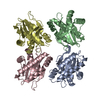
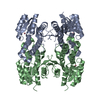
- Assembly
| Deposited unit | 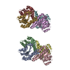
| ||||||||
|---|---|---|---|---|---|---|---|---|---|
| 1 | 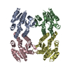
| ||||||||
| 2 | 
| ||||||||
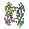
| Unit cell |
|
-Components
| #1: Protein | Mass: 26419.887 Da / Num. of mol.: 8 Source method: isolated from a genetically manipulated source Source: (gene. exp.) Corynebacterium glutamicum (bacteria) / Gene: Cgl2444, cg2685 / Plasmid: modified pET26 / Production host: #2: Water | ChemComp-HOH / |  Mass: 18.015 Da / Num. of mol.: 127 / Source method: isolated from a natural source / Formula: H2O Mass: 18.015 Da / Num. of mol.: 127 / Source method: isolated from a natural source / Formula: H2O |
|---|
-Experimental details
-Experiment
| Experiment | Method: X-RAY DIFFRACTION / Number of used crystals: 1 |
|---|
- Sample preparation
| Crystal | Density Matthews: 2.2 Å3/Da / Density % sol: 44.18 % |
|---|---|
| Crystal grow | Temperature: 294 K / Method: vapor diffusion / pH: 6 Details: 100mM MES pH 6.0, 27% PEG MME 5K, 200mM ammonium sulfate, Vapor diffusion, temperature 294K |
-Data collection
| Diffraction | Mean temperature: 100 K |
|---|---|
| Diffraction source | Source: SYNCHROTRON / Site: APS  / Beamline: 31-ID / Wavelength: 0.97958 Å / Beamline: 31-ID / Wavelength: 0.97958 Å |
| Detector | Type: MAR CCD 165 mm / Detector: CCD / Date: Jul 2, 2008 |
| Radiation | Monochromator: diamond / Protocol: SINGLE WAVELENGTH / Monochromatic (M) / Laue (L): M / Scattering type: x-ray |
| Radiation wavelength | Wavelength: 0.97958 Å / Relative weight: 1 |
| Reflection | Resolution: 2.4→29.54 Å / Num. all: 72534 / Num. obs: 72461 / % possible obs: 99.9 % / Observed criterion σ(F): 0 / Observed criterion σ(I): 0 / Redundancy: 5.5 % / Biso Wilson estimate: 40.3 Å2 / Rmerge(I) obs: 0.096 / Rsym value: 0.096 / Net I/σ(I): 13.3 |
| Reflection shell | Resolution: 2.4→2.53 Å / Redundancy: 5.3 % / Rmerge(I) obs: 0.397 / Mean I/σ(I) obs: 3.7 / Num. measured all: 55025 / Num. unique all: 10458 / Rsym value: 0.397 / % possible all: 99.8 |
- Processing
| Software |
| |||||||||||||||||||||||||||||||||||||||||||||||||||||||||||||||||
|---|---|---|---|---|---|---|---|---|---|---|---|---|---|---|---|---|---|---|---|---|---|---|---|---|---|---|---|---|---|---|---|---|---|---|---|---|---|---|---|---|---|---|---|---|---|---|---|---|---|---|---|---|---|---|---|---|---|---|---|---|---|---|---|---|---|---|
| Refinement | Method to determine structure: SAD / Resolution: 2.4→20 Å / Cor.coef. Fo:Fc: 0.929 / Cor.coef. Fo:Fc free: 0.906 / WRfactor Rfree: 0.271 / WRfactor Rwork: 0.225 / Occupancy max: 1 / Occupancy min: 0.5 / FOM work R set: 0.77 / SU B: 8.167 / SU ML: 0.195 / SU R Cruickshank DPI: 0.367 / SU Rfree: 0.268 / Cross valid method: THROUGHOUT / σ(F): 0 / σ(I): 0 / ESU R: 0.367 / ESU R Free: 0.268 / Stereochemistry target values: MAXIMUM LIKELIHOOD / Details: HYDROGENS HAVE BEEN ADDED IN THE RIDING POSITIONS
| |||||||||||||||||||||||||||||||||||||||||||||||||||||||||||||||||
| Solvent computation | Ion probe radii: 0.8 Å / Shrinkage radii: 0.8 Å / VDW probe radii: 1.2 Å / Solvent model: BABINET MODEL WITH MASK | |||||||||||||||||||||||||||||||||||||||||||||||||||||||||||||||||
| Displacement parameters | Biso max: 104.74 Å2 / Biso mean: 45.14 Å2 / Biso min: 20.97 Å2
| |||||||||||||||||||||||||||||||||||||||||||||||||||||||||||||||||
| Refinement step | Cycle: LAST / Resolution: 2.4→20 Å
| |||||||||||||||||||||||||||||||||||||||||||||||||||||||||||||||||
| Refine LS restraints |
| |||||||||||||||||||||||||||||||||||||||||||||||||||||||||||||||||
| LS refinement shell | Resolution: 2.4→2.461 Å / Total num. of bins used: 20
|
