 Movie
Movie Controller

[English] 日本語
 Yorodumi
- PDB-3e3u: Crystal structure of Mycobacterium tuberculosis peptide deformyla... -
+ Open data

- Basic information
| Entry | Database: PDB / ID: 3e3u | ||||||
|---|---|---|---|---|---|---|---|
| Title | Crystal structure of Mycobacterium tuberculosis peptide deformylase in complex with inhibitor | ||||||
 Components Components | Peptide deformylase | ||||||
 Keywords Keywords | HYDROLASE / metallo-enzyme / Iron / Metal-binding / Protein biosynthesis | ||||||
| Function / homology |  Function and homology information Function and homology informationpeptide deformylase / peptide deformylase activity / translation / metal ion binding Similarity search - Function | ||||||
| Biological species |   Mycobacterium tuberculosis (bacteria) Mycobacterium tuberculosis (bacteria) | ||||||
| Method |  X-RAY DIFFRACTION / FOURIER SYNTHESIS / Resolution: 1.56 Å X-RAY DIFFRACTION / FOURIER SYNTHESIS / Resolution: 1.56 Å | ||||||
 Authors Authors | Meng, W. / Xu, M. / Pan, S. / Koehn, J. | ||||||
 Citation Citation | Journal: Bioorg.Med.Chem.Lett. / Year: 2008 Title: Peptide deformylase inhibitors of Mycobacterium tuberculosis: synthesis, structural investigations, and biological results. Authors: Pichota, A. / Duraiswamy, J. / Yin, Z. / Keller, T.H. / Alam, J. / Liung, S. / Lee, G. / Ding, M. / Wang, G. / Chan, W.L. / Schreiber, M. / Ma, I. / Beer, D. / Ngew, X. / Mukherjee, K. / ...Authors: Pichota, A. / Duraiswamy, J. / Yin, Z. / Keller, T.H. / Alam, J. / Liung, S. / Lee, G. / Ding, M. / Wang, G. / Chan, W.L. / Schreiber, M. / Ma, I. / Beer, D. / Ngew, X. / Mukherjee, K. / Nanjundappa, M. / Teo, J.W. / Thayalan, P. / Yap, A. / Dick, T. / Meng, W. / Xu, M. / Koehn, J. / Pan, S.H. / Clark, K. / Xie, X. / Shoen, C. / Cynamon, M. | ||||||
| History |
|
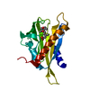
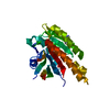
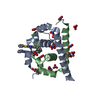
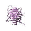
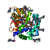
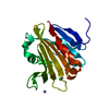
- Structure visualization
| Structure viewer | Molecule: MolmilJmol/JSmol |
|---|
- Downloads & links
-Download
| PDBx/mmCIF format | 3e3u.cif.gz | 57.8 KB | Display | PDBx/mmCIF format |
|---|---|---|---|---|
| PDB format | pdb3e3u.ent.gz | 41.4 KB | Display | PDB format |
| PDBx/mmJSON format | 3e3u.json.gz | Tree view | PDBx/mmJSON format | |
| Others |  Other downloads Other downloads |
-Validation report
| Arichive directory | https://data.pdbj.org/pub/pdb/validation_reports/e3/3e3uftp://data.pdbj.org/pub/pdb/validation_reports/e3/3e3u | HTTPS FTP |
|---|
-Related structure data
| Similar structure data |
|---|


-Links
 PDBj
PDBj- Assembly
| Deposited unit | 
| ||||||||
|---|---|---|---|---|---|---|---|---|---|
| 1 |
| ||||||||
| Unit cell |
|
-Components
| #1: Protein | Mass: 20959.689 Da / Num. of mol.: 1 Source method: isolated from a genetically manipulated source Source: (gene. exp.) Mycobacterium tuberculosis (bacteria) / Gene: def / Plasmid: pET24a / Production host: References: UniProt: P96275, UniProt: P9WIJ3*PLUS, peptide deformylase | ||||
|---|---|---|---|---|---|
| #2: Chemical | ChemComp-NI /   Mass: 58.693 Da / Num. of mol.: 1 / Source method: obtained synthetically / Formula: Ni Mass: 58.693 Da / Num. of mol.: 1 / Source method: obtained synthetically / Formula: Ni | ||||
| #3: Chemical | 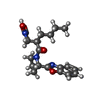  Mass: 359.420 Da / Num. of mol.: 3 / Source method: obtained synthetically / Formula: C19H25N3O4 Mass: 359.420 Da / Num. of mol.: 3 / Source method: obtained synthetically / Formula: C19H25N3O4#4: Water | ChemComp-HOH / |  Mass: 18.015 Da / Num. of mol.: 259 / Source method: isolated from a natural source / Formula: H2O Mass: 18.015 Da / Num. of mol.: 259 / Source method: isolated from a natural source / Formula: H2ONonpolymer details | BIOLOGICAL | |
-Experimental details
-Experiment
| Experiment | Method: X-RAY DIFFRACTION / Number of used crystals: 1 |
|---|
- Sample preparation
| Crystal | Density Matthews: 2.64 Å3/Da / Density % sol: 53.44 % |
|---|---|
| Crystal grow | Temperature: 298 K / Method: vapor diffusion, hanging drop / pH: 7.5 Details: 2.0 M Ammonium sulfate, Tris-HCl pH 7.5, 5mM Compound 16a, VAPOR DIFFUSION, HANGING DROP, temperature 298K |
-Data collection
| Diffraction | Mean temperature: 100 K |
|---|---|
| Diffraction source | Source: ROTATING ANODE / Type: RIGAKU FR-E SUPERBRIGHT / Wavelength: 1.5418 Å |
| Detector | Type: RIGAKU RAXIS IV / Detector: IMAGE PLATE / Date: Mar 23, 2005 / Details: Multi-layer mirrors |
| Radiation | Monochromator: Ni FILTER / Protocol: SINGLE WAVELENGTH / Monochromatic (M) / Laue (L): M / Scattering type: x-ray |
| Radiation wavelength | Wavelength: 1.5418 Å / Relative weight: 1 |
| Reflection | Resolution: 1.56→50.32 Å / Num. all: 32726 / Num. obs: 32726 / % possible obs: 99.6 % / Observed criterion σ(I): -3 / Redundancy: 3.5 % / Rmerge(I) obs: 0.062 / Net I/σ(I): 30.6 |
| Reflection shell | Resolution: 1.56→1.62 Å / Redundancy: 3.1 % / Rmerge(I) obs: 0.475 / Mean I/σ(I) obs: 2.9 / Num. unique all: 3027 / % possible all: 97.2 |
- Processing
| Software |
| ||||||||||||||||||||||||||||||||
|---|---|---|---|---|---|---|---|---|---|---|---|---|---|---|---|---|---|---|---|---|---|---|---|---|---|---|---|---|---|---|---|---|---|
| Refinement | Method to determine structure: FOURIER SYNTHESIS / Resolution: 1.56→50.32 Å / Cor.coef. Fo:Fc: 0.967 / Cor.coef. Fo:Fc free: 0.955 / Occupancy max: 1 / Occupancy min: 0.5 / SU B: 1.418 / SU ML: 0.052 / Cross valid method: THROUGHOUT / σ(F): 0 / ESU R: 0.077 / ESU R Free: 0.08 / Stereochemistry target values: MAXIMUM LIKELIHOOD
| ||||||||||||||||||||||||||||||||
| Solvent computation | Ion probe radii: 0.8 Å / Shrinkage radii: 0.8 Å / VDW probe radii: 1.2 Å / Solvent model: MASK | ||||||||||||||||||||||||||||||||
| Displacement parameters | Biso max: 61 Å2 / Biso mean: 18.589 Å2 / Biso min: 9.58 Å2
| ||||||||||||||||||||||||||||||||
| Refinement step | Cycle: LAST / Resolution: 1.56→50.32 Å
| ||||||||||||||||||||||||||||||||
| Refine LS restraints |
| ||||||||||||||||||||||||||||||||
| LS refinement shell | Resolution: 1.56→1.598 Å / Total num. of bins used: 20
|
