Mass: 18.015 Da / Num. of mol.: 300 / Source method: isolated from a natural source / Formula: H2O
-
Details
Has protein modification
Y
Nonpolymer details
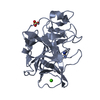
THE REFERENCE OF I3C IS ACTA CRYSTALLOGRAPHICA SECTION E(2008) E64, O1286 TITLE: 5-AMINO-2,4,6- ...THE REFERENCE OF I3C IS ACTA CRYSTALLOGRAPHICA SECTION E(2008) E64, O1286 TITLE: 5-AMINO-2,4,6-TRIIODOISOPHTHALIC ACID MONOHYDRATE AUTHOR: T. BECK AND G.M. SHELDRICK
Sequence details
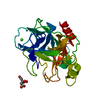
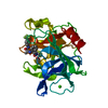
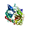
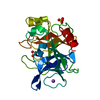
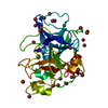
THIS SEQUENCE IS FROM REFERENCE 3 IN UNP DATABASE, P00772. BIOCHEM. J. 131:643-675(1973) SHOTTON D. ...THIS SEQUENCE IS FROM REFERENCE 3 IN UNP DATABASE, P00772. BIOCHEM. J. 131:643-675(1973) SHOTTON D.M., HARTLEY B.S. EVIDENCE FOR THE AMINO ACID SEQUENCE OF PORCINE PANCREATIC ELASTASE
-
Experimental details
-
Experiment
Experiment
Method: X-RAY DIFFRACTION / Number of used crystals: 1
-
Sample preparation
Crystal
Density Matthews: 2.083245 Å3/Da / Density % sol: 40.96 %
Crystal grow
Temperature: 277 K / Method: vapor diffusion, sitting drop / pH: 8 Details: 0.1M HEPES, 0.6M sodium sulfate, pH 8.0, VAPOR DIFFUSION, SITTING DROP, temperature 277K
-
Data collection
Diffraction
Mean temperature: 100 K
Diffraction source
Source: ROTATING ANODE / Type: MACSCIENCE / Wavelength: 1.54178 Å
Detector
Type: BRUKER SMART 6000 / Detector: CCD / Date: Mar 28, 2008 / Details: MULTI-LAYER INCOATEC OPTICS
Radiation
Monochromator: MULTI-LAYER INCOATEC OPTICS / Protocol: SINGLE WAVELENGTH / Monochromatic (M) / Laue (L): M / Scattering type: x-ray
In the structure databanks used in Yorodumi, some data are registered as the other names, "COVID-19 virus" and "2019-nCoV". Here are the details of the virus and the list of structure data.
Jan 31, 2019. EMDB accession codes are about to change! (news from PDBe EMDB page)
EMDB accession codes are about to change! (news from PDBe EMDB page)
The allocation of 4 digits for EMDB accession codes will soon come to an end. Whilst these codes will remain in use, new EMDB accession codes will include an additional digit and will expand incrementally as the available range of codes is exhausted. The current 4-digit format prefixed with “EMD-” (i.e. EMD-XXXX) will advance to a 5-digit format (i.e. EMD-XXXXX), and so on. It is currently estimated that the 4-digit codes will be depleted around Spring 2019, at which point the 5-digit format will come into force.
The EM Navigator/Yorodumi systems omit the EMD- prefix.
Related info.:Q: What is EMD? / ID/Accession-code notation in Yorodumi/EM Navigator
Yorodumi is a browser for structure data from EMDB, PDB, SASBDB, etc.
This page is also the successor to EM Navigator detail page, and also detail information page/front-end page for Omokage search.
The word "yorodu" (or yorozu) is an old Japanese word meaning "ten thousand". "mi" (miru) is to see.
Related info.:EMDB / PDB / SASBDB / Comparison of 3 databanks / Yorodumi Search / Aug 31, 2016. New EM Navigator & Yorodumi / Yorodumi Papers / Jmol/JSmol / Function and homology information / Changes in new EM Navigator and Yorodumi
 Movie
Movie Controller

 Yorodumi
 Open data
 Basic information
 Components
 Keywords
 Function and homology information
 X-RAY DIFFRACTION /
  Authors
 Citation
 Structure visualization
 Downloads & links
 Other downloads
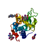


 PDBj
PDBj
 Assembly


 Mass: 96.063 Da / Num. of mol.: 2 / Source method: obtained synthetically / Formula: SO4
 Mass: 22.990 Da / Num. of mol.: 1 / Source method: obtained synthetically / Formula: Na
 Mass: 126.904 Da / Num. of mol.: 1 / Source method: obtained synthetically / Formula: I
 Mass: 558.835 Da / Num. of mol.: 4 / Source method: obtained synthetically / Formula: C8H4I3NO4
 Sample preparation
 Processing