 Movie
Movie Controller

[English] 日本語
 Yorodumi
- PDB-1m9u: Crystal Structure of Earthworm Fibrinolytic Enzyme Component A fr... -
+ Open data

- Basic information
| Entry | Database: PDB / ID: 1m9u | |||||||||
|---|---|---|---|---|---|---|---|---|---|---|
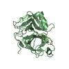
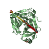
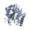
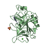
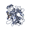
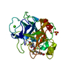
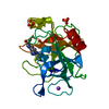
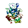
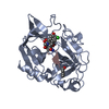
| Title | Crystal Structure of Earthworm Fibrinolytic Enzyme Component A from Eisenia fetida | |||||||||
 Components Components | Earthworm Fibrinolytic Enzyme | |||||||||
 Keywords Keywords | HYDROLASE / SERINE PROTEASE (ELASTASE-LIKE) / FIBRINOLYTIC ENZYME | |||||||||
| Function / homology |  Function and homology information Function and homology information | |||||||||
| Biological species |  | |||||||||
| Method |  X-RAY DIFFRACTION / MIR / Resolution: 2.3 Å X-RAY DIFFRACTION / MIR / Resolution: 2.3 Å | |||||||||
 Authors Authors | Chang, W. / Liang, D. / Tang, Y. | |||||||||
 Citation Citation | Journal: J.Mol.Biol. / Year: 2002 Title: Crystal structure of earthworm fibrinolytic enzyme component a: revealing the structural determinants of its dual fibrinolytic activity. Authors: Tang, Y. / Liang, D. / Jiang, T. / Zhang, J. / Gui, L. / Chang, W. #1: Journal: Acta Crystallogr.,Sect.D / Year: 2000Title: Crystallization and Preliminary X-ray Analysis of Earthworm Fibrinolytic Enzyme Component A from Eisenia fetida Authors: Tang, Y. / Zhang, J. / Gui, L. / Wu, C. / Fan, R. / Chang, W. / Liang, D. #2: Journal: ENZYMES 3RD ED. / Year: 1971Title: Pancreatic Elastase Authors: Hartley, B.S. / Shotton, D.M. | |||||||||
| History |
|
- Structure visualization
| Structure viewer | Molecule: MolmilJmol/JSmol |
|---|
- Downloads & links
-Download
| PDBx/mmCIF format | 1m9u.cif.gz | 142.1 KB | Display | PDBx/mmCIF format |
|---|---|---|---|---|
| PDB format | pdb1m9u.ent.gz | 111.9 KB | Display | PDB format |
| PDBx/mmJSON format | 1m9u.json.gz | Tree view | PDBx/mmJSON format | |
| Others |  Other downloads Other downloads |
-Validation report
| Arichive directory | https://data.pdbj.org/pub/pdb/validation_reports/m9/1m9uftp://data.pdbj.org/pub/pdb/validation_reports/m9/1m9u | HTTPS FTP |
|---|
-Related structure data
| Similar structure data |
|---|

-Links
 PDBj
PDBj- Assembly
| Deposited unit | 
| ||||||||
|---|---|---|---|---|---|---|---|---|---|
| 1 | 
| ||||||||
| 2 | 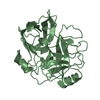
| ||||||||
| 3 | 
| ||||||||
| Unit cell |
|
-Components
| #1: Protein | Mass: 24661.951 Da / Num. of mol.: 3 / Fragment: component A / Source method: isolated from a natural source / Source: (natural) References: UniProt: Q8MX72, Hydrolases; Acting on peptide bonds (peptidases); Serine endopeptidases #2: Water | ChemComp-HOH / |  Mass: 18.015 Da / Num. of mol.: 278 / Source method: isolated from a natural source / Formula: H2O Mass: 18.015 Da / Num. of mol.: 278 / Source method: isolated from a natural source / Formula: H2OHas protein modification | Y | |
|---|
-Experimental details
-Experiment
| Experiment | Method: X-RAY DIFFRACTION / Number of used crystals: 1 |
|---|
- Sample preparation
| Crystal | Density Matthews: 2.24 Å3/Da / Density % sol: 45 % |
|---|---|
| Crystal grow | Temperature: 291 K / Method: vapor diffusion, hanging drop / pH: 7.2 Details: Ammonium Sulfate, PEG400, MOPS, pH 7.2, VAPOR DIFFUSION, HANGING DROP, temperature 291K |
| Crystal grow | *PLUS Details: Otwinowski, Z., (1997) Methods Enzymol., 276, 307. |
-Data collection
| Diffraction | Mean temperature: 291 K |
|---|---|
| Diffraction source | Source: SEALED TUBE / Type: CONVENTIONAL Cu / Wavelength: 1.5418 Å |
| Detector | Type: MARRESEARCH / Detector: IMAGE PLATE / Date: Mar 15, 2000 / Details: mirrors |
| Radiation | Monochromator: graphite / Protocol: SINGLE WAVELENGTH / Monochromatic (M) / Laue (L): M / Scattering type: x-ray |
| Radiation wavelength | Wavelength: 1.5418 Å / Relative weight: 1 |
| Reflection | Resolution: 2.3→9 Å / Num. all: 29792 / Num. obs: 29703 / % possible obs: 99.7 % / Observed criterion σ(F): 0 / Observed criterion σ(I): -3 / Redundancy: 16 % / Biso Wilson estimate: 32.99 Å2 / Rmerge(I) obs: 0.156 / Net I/σ(I): 16.2 |
| Reflection shell | Resolution: 2.3→2.35 Å / Redundancy: 5.3 % / Rmerge(I) obs: 0.586 / Mean I/σ(I) obs: 2.4 / % possible all: 98.9 |
| Reflection | *PLUS Rmerge(I) obs: 0.162 |
| Reflection shell | *PLUS Highest resolution: 2.3 Å / % possible obs: 98.9 % |
- Processing
| Software |
| |||||||||||||||||||||||||
|---|---|---|---|---|---|---|---|---|---|---|---|---|---|---|---|---|---|---|---|---|---|---|---|---|---|---|
| Refinement | Method to determine structure: MIR / Resolution: 2.3→9 Å / Isotropic thermal model: Isotropic / Cross valid method: THROUGHOUT / σ(F): 0 / Stereochemistry target values: Engh & Huber / Details: NCS RESTRAINED
| |||||||||||||||||||||||||
| Displacement parameters | Biso mean: 19.2 Å2 | |||||||||||||||||||||||||
| Refinement step | Cycle: LAST / Resolution: 2.3→9 Å
| |||||||||||||||||||||||||
| Refine LS restraints |
| |||||||||||||||||||||||||
| LS refinement shell | Resolution: 2.3→2.4 Å / Total num. of bins used: 8
| |||||||||||||||||||||||||
| Refinement | *PLUS Highest resolution: 2.3 Å / % reflection Rfree: 8 % | |||||||||||||||||||||||||
| Solvent computation | *PLUS | |||||||||||||||||||||||||
| Displacement parameters | *PLUS | |||||||||||||||||||||||||
| Refine LS restraints | *PLUS
|
