 ムービー
ムービー コントローラー


+ データを開く

- 基本情報
基本情報
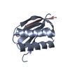
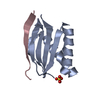
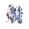
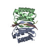
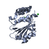
| 登録情報 | データベース: PDB / ID: 3e2b | |||||||||
|---|---|---|---|---|---|---|---|---|---|---|
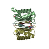
| タイトル | Crystal structure of Dynein Light chain LC8 in complex with a peptide derived from Swallow | |||||||||
 要素 要素 |
| |||||||||
 キーワード キーワード | TRANSPORT PROTEIN / protein-peptide complex / Cytoplasm / Dynein / Microtubule / Motor protein | |||||||||
| 機能・相同性 |  機能・相同性情報 機能・相同性情報pole plasm mRNA localization / bicoid mRNA localization / spermatid nucleus elongation / regulation of pole plasm oskar mRNA localization / chaeta morphogenesis / Macroautophagy / Aggrephagy / positive regulation of neuron remodeling / wing disc development / HSP90 chaperone cycle for steroid hormone receptors (SHR) in the presence of ligand ...pole plasm mRNA localization / bicoid mRNA localization / spermatid nucleus elongation / regulation of pole plasm oskar mRNA localization / chaeta morphogenesis / Macroautophagy / Aggrephagy / positive regulation of neuron remodeling / wing disc development / HSP90 chaperone cycle for steroid hormone receptors (SHR) in the presence of ligand / COPI-mediated anterograde transport / COPI-independent Golgi-to-ER retrograde traffic / chaeta development / sperm individualization / imaginal disc-derived wing morphogenesis / transposable element silencing by piRNA-mediated heterochromatin formation / RNA polymerase II transcription repressor complex / microtubule anchoring at centrosome / anterior/posterior axis specification, embryo / Neutrophil degranulation / dynein complex / cytoplasmic dynein complex / dynein light intermediate chain binding / oogenesis / dynein intermediate chain binding / dynein complex binding / establishment of mitotic spindle orientation / actin filament bundle assembly / centriole / actin filament organization / disordered domain specific binding / transcription corepressor activity / spermatogenesis / microtubule / cell division / mRNA binding / protein homodimerization activity / protein-containing complex / nucleus / cytoplasm / cytosol 類似検索 - 分子機能 | |||||||||
| 生物種 |  | |||||||||
| 手法 |  X線回折 / シンクロトロン / 分子置換 / 解像度: 2 Å X線回折 / シンクロトロン / 分子置換 / 解像度: 2 Å | |||||||||
 データ登録者 データ登録者 | Benison, G. / Barbar, E. / Karplus, P.A. | |||||||||
 引用 引用 | ジャーナル: J.Mol.Biol. / 年: 2008 タイトル: The interplay of ligand binding and quaternary structure in the diverse interactions of dynein light chain LC8. 著者: Benison, G. / Karplus, P.A. / Barbar, E. #1: ジャーナル: J.Mol.Biol. / 年: 2007タイトル: Structure and dynamics of LC8 complexes with KXTQT-motif peptides: swallow and dynein intermediate chain compete for a common site. 著者: Benison, G. / Karplus, P.A. / Barbar, E. | |||||||||
| 履歴 |
|
- 構造の表示
構造の表示
| 構造ビューア | 分子: MolmilJmol/JSmol |
|---|
- ダウンロードとリンク
-ダウンロード
| PDBx/mmCIF形式 | 3e2b.cif.gz | 56.3 KB | 表示 | PDBx/mmCIF形式 |
|---|---|---|---|---|
| PDB形式 | pdb3e2b.ent.gz | 41 KB | 表示 | PDB形式 |
| PDBx/mmJSON形式 | 3e2b.json.gz | ツリー表示 | PDBx/mmJSON形式 | |
| その他 |  その他のダウンロード その他のダウンロード |
-検証レポート
| 文書・要旨 | 3e2b_validation.pdf.gz | 431.9 KB | 表示 | wwPDB検証レポート |
|---|---|---|---|---|
| 文書・詳細版 | 3e2b_full_validation.pdf.gz | 432.7 KB | 表示 | |
| XML形式データ | 3e2b_validation.xml.gz | 7 KB | 表示 | |
| CIF形式データ | 3e2b_validation.cif.gz | 8.6 KB | 表示 | |
| アーカイブディレクトリ | https://data.pdbj.org/pub/pdb/validation_reports/e2/3e2bftp://data.pdbj.org/pub/pdb/validation_reports/e2/3e2b | HTTPS FTP |
-関連構造データ


-リンク
 PDBj
PDBj


- 集合体
集合体
| 登録構造単位 | 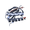
| ||||||||
|---|---|---|---|---|---|---|---|---|---|
| 1 | 
| ||||||||
| 単位格子 |
|
-要素
| #1: タンパク質 | 分子量: 10388.849 Da / 分子数: 1 / 由来タイプ: 組換発現 由来: (組換発現) 遺伝子: ctp, Cdlc1, ddlc1, CG6998 / 発現宿主:  |
|---|---|
| #2: タンパク質・ペプチド | 分子量: 1783.980 Da / 分子数: 1 / 由来タイプ: 合成 詳細: The peptide was chemically synthesized. The sequence of the peptide can be naturally found in Drosophila melanogaster (Fruit fly). 参照: UniProt: P40688 |
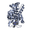
| #3: 化合物 | ChemComp-ACT /   分子量: 59.044 Da / 分子数: 1 / 由来タイプ: 合成 / 式: C2H3O2 分子量: 59.044 Da / 分子数: 1 / 由来タイプ: 合成 / 式: C2H3O2 |
| #4: 水 | ChemComp-HOH /  分子量: 18.015 Da / 分子数: 61 / 由来タイプ: 天然 / 式: H2O 分子量: 18.015 Da / 分子数: 61 / 由来タイプ: 天然 / 式: H2O |
-実験情報
-実験
| 実験 | 手法: X線回折 / 使用した結晶の数: 1 |
|---|
- 試料調製
試料調製
| 結晶 | マシュー密度: 2.36 Å3/Da / 溶媒含有率: 47.77 % |
|---|---|
| 結晶化 | 温度: 298 K / 手法: 蒸気拡散法, ハンギングドロップ法 / pH: 5.5 詳細: 0.2M sodium potassium tartrate, 0.1M sodium citrate, 2.0M ammonium sulfate, pH 5.5, VAPOR DIFFUSION, HANGING DROP, temperature 298K |
-データ収集
| 回折 | 平均測定温度: 77 K |
|---|---|
| 放射光源 | 由来: シンクロトロン / サイト: ALS  / ビームライン: 8.2.1 / 波長: 0.979 Å / ビームライン: 8.2.1 / 波長: 0.979 Å |
| 検出器 | 検出器: AREA DETECTOR / 日付: 2008年2月27日 |
| 放射 | プロトコル: SINGLE WAVELENGTH / 単色(M)・ラウエ(L): M / 散乱光タイプ: x-ray |
| 放射波長 | 波長: 0.979 Å / 相対比: 1 |
| 反射 | 解像度: 1.9→40.7 Å / Num. all: 10184 / Num. obs: 10184 / % possible obs: 100 % / 冗長度: 13.7 % / Biso Wilson estimate: 28.4 Å2 / Rsym value: 0.07 / Net I/σ(I): 23.5 |
| 反射 シェル | 解像度: 1.9→2 Å / 冗長度: 14.3 % / Mean I/σ(I) obs: 5.1 / Num. unique all: 1436 / Rsym value: 0.63 / % possible all: 100 |
- 解析
解析
| ソフトウェア |
| ||||||||||||||||||||||||||||||||||||||||||||||||||||||||||||||||||||||||||||||||||||||||||
|---|---|---|---|---|---|---|---|---|---|---|---|---|---|---|---|---|---|---|---|---|---|---|---|---|---|---|---|---|---|---|---|---|---|---|---|---|---|---|---|---|---|---|---|---|---|---|---|---|---|---|---|---|---|---|---|---|---|---|---|---|---|---|---|---|---|---|---|---|---|---|---|---|---|---|---|---|---|---|---|---|---|---|---|---|---|---|---|---|---|---|---|
| 精密化 | 構造決定の手法: 分子置換 開始モデル: pdb entry 2p1k  2p1k 解像度: 2→35.81 Å / Cor.coef. Fo:Fc: 0.95 / Cor.coef. Fo:Fc free: 0.925 / SU B: 11.277 / SU ML: 0.154 / 交差検証法: THROUGHOUT / ESU R: 0.2 / ESU R Free: 0.183 / 立体化学のターゲット値: MAXIMUM LIKELIHOOD / 詳細: HYDROGENS HAVE BEEN ADDED IN THE RIDING POSITIONS
| ||||||||||||||||||||||||||||||||||||||||||||||||||||||||||||||||||||||||||||||||||||||||||
| 溶媒の処理 | イオンプローブ半径: 0.8 Å / 減衰半径: 0.8 Å / VDWプローブ半径: 1.4 Å / 溶媒モデル: BABINET MODEL WITH MASK | ||||||||||||||||||||||||||||||||||||||||||||||||||||||||||||||||||||||||||||||||||||||||||
| 原子変位パラメータ | Biso mean: 34.422 Å2
| ||||||||||||||||||||||||||||||||||||||||||||||||||||||||||||||||||||||||||||||||||||||||||
| 精密化ステップ | サイクル: LAST / 解像度: 2→35.81 Å
| ||||||||||||||||||||||||||||||||||||||||||||||||||||||||||||||||||||||||||||||||||||||||||
| 拘束条件 |
| ||||||||||||||||||||||||||||||||||||||||||||||||||||||||||||||||||||||||||||||||||||||||||
| LS精密化 シェル | 解像度: 2→2.052 Å / Total num. of bins used: 20
|
