 Movie
Movie Controller

[English] 日本語
 Yorodumi
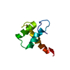
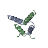
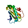
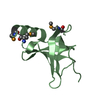
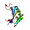
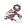
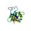
- PDB-3e0e: Crystal structure of a domain of replication protein A from Metha... -
+ Open data

- Basic information
| Entry | Database: PDB / ID: 3e0e | ||||||
|---|---|---|---|---|---|---|---|
| Title | Crystal structure of a domain of replication protein A from Methanococcus maripaludis. NorthEast Structural Genomics targe MrR110B | ||||||
 Components Components | Replication protein A | ||||||
 Keywords Keywords | REPLICATION / Replication protein A / Structural Genomics / PSI-2 / Protein Structure Initiative / Northeast Structural Genomics Consortium / NESG | ||||||
| Function / homology |  Function and homology information Function and homology informationresponse to ionizing radiation / double-strand break repair via homologous recombination / DNA binding Similarity search - Function | ||||||
| Biological species |  Methanococcus maripaludis (archaea) Methanococcus maripaludis (archaea) | ||||||
| Method |  X-RAY DIFFRACTION / SYNCHROTRON / SAD / Resolution: 1.6 Å X-RAY DIFFRACTION / SYNCHROTRON / SAD / Resolution: 1.6 Å | ||||||
 Authors Authors | Seetharaman, J. / Chen, Y. / Wang, H. / Janjua, H. / Foote, E.L. / Xiao, R. / Nair, R. / Everett, J.K. / Acton, T.B. / Rost, B. ...Seetharaman, J. / Chen, Y. / Wang, H. / Janjua, H. / Foote, E.L. / Xiao, R. / Nair, R. / Everett, J.K. / Acton, T.B. / Rost, B. / Montelione, G.T. / Hunt, J.F. / Tong, L. / Northeast Structural Genomics Consortium (NESG) | ||||||
 Citation Citation | Journal: To be Published Title: Crystal structure of a domain of replication protein A from Methanococcus maripaludis. NorthEast Structural Genomics targe MrR110B Authors: Seetharaman, J. / Chen, Y. / Wang, H. / Janjua, H. / Foote, E.L. / Xiao, R. / Nair, R. / Everett, J.K. / Acton, T.B. / Rost, B. / Montelione, G.T. / Hunt, J.F. / Tong, L. | ||||||
| History |
|
- Structure visualization
| Structure viewer | Molecule: MolmilJmol/JSmol |
|---|
- Downloads & links
-Download
| PDBx/mmCIF format | 3e0e.cif.gz | 32.2 KB | Display | PDBx/mmCIF format |
|---|---|---|---|---|
| PDB format | pdb3e0e.ent.gz | 20.7 KB | Display | PDB format |
| PDBx/mmJSON format | 3e0e.json.gz | Tree view | PDBx/mmJSON format | |
| Others |  Other downloads Other downloads |
-Validation report
| Arichive directory | https://data.pdbj.org/pub/pdb/validation_reports/e0/3e0eftp://data.pdbj.org/pub/pdb/validation_reports/e0/3e0e | HTTPS FTP |
|---|
-Related structure data
| Similar structure data | |
|---|---|
| Other databases |


-Links
 PDBj
PDBj

- Assembly
| Deposited unit | 
| ||||||||
|---|---|---|---|---|---|---|---|---|---|
| 1 |
| ||||||||
| Unit cell |
|
-Components
| #1: Protein | Mass: 10796.943 Da / Num. of mol.: 1 / Fragment: residues 173-267 Source method: isolated from a genetically manipulated source Source: (gene. exp.) Methanococcus maripaludis (archaea) / Gene: rpa, MMP1032 / Plasmid: PET 21 / Production host:  |
|---|---|
| #2: Water | ChemComp-HOH /  Mass: 18.015 Da / Num. of mol.: 104 / Source method: isolated from a natural source / Formula: H2O Mass: 18.015 Da / Num. of mol.: 104 / Source method: isolated from a natural source / Formula: H2O |
| Has protein modification | Y |
-Experimental details
-Experiment
| Experiment | Method: X-RAY DIFFRACTION / Number of used crystals: 1 |
|---|
- Sample preparation
| Crystal | Density Matthews: 1.81 Å3/Da / Density % sol: 31.87 % |
|---|---|
| Crystal grow | Temperature: 293 K / pH: 5 Details: Sodium chloride 0.1 M, Sodium Acetate 0.1 M, PEG 4000 40%, pH 5, Micro batch under oil method, temperature 293K |
-Data collection
| Diffraction |
| |||||||||||||||
|---|---|---|---|---|---|---|---|---|---|---|---|---|---|---|---|---|
| Diffraction source |
| |||||||||||||||
| Detector | Type: ADSC QUANTUM 210 / Detector: CCD / Date: Jun 29, 2008 / Details: Mirrors | |||||||||||||||
| Radiation | Protocol: SINGLE WAVELENGTH / Monochromatic (M) / Laue (L): M / Scattering type: x-ray | |||||||||||||||
| Radiation wavelength | Wavelength: 0.979 Å / Relative weight: 1 | |||||||||||||||
| Reflection | Resolution: 1.6→50 Å / Num. obs: 19963 / % possible obs: 99.7 % / Observed criterion σ(F): 0 / Observed criterion σ(I): 0 / Redundancy: 21.4 % / Biso Wilson estimate: 17.5 Å2 / Rmerge(I) obs: 0.048 / Rsym value: 0.044 | |||||||||||||||
| Reflection shell | Resolution: 1.6→1.66 Å / Redundancy: 3.3 % / Rmerge(I) obs: 0.225 / Mean I/σ(I) obs: 19.5 / Num. unique all: 1987 / Rsym value: 0.213 / % possible all: 98.7 |

- Processing
| Software |
| ||||||||||||||||||||
|---|---|---|---|---|---|---|---|---|---|---|---|---|---|---|---|---|---|---|---|---|---|
| Refinement | Method to determine structure: SAD / Resolution: 1.6→39.28 Å / Rfactor Rfree error: 0.008 / Data cutoff high absF: 146558 / Data cutoff low absF: 0 / Isotropic thermal model: RESTRAINED / Cross valid method: THROUGHOUT / σ(F): 0 / Stereochemistry target values: Engh & Huber / Details: BULK SOLVENT MODEL USED
| ||||||||||||||||||||
| Solvent computation | Solvent model: FLAT MODEL / Bsol: 61.0129 Å2 / ksol: 0.4 e/Å3 | ||||||||||||||||||||
| Displacement parameters | Biso mean: 18.2 Å2
| ||||||||||||||||||||
| Refine analyze |
| ||||||||||||||||||||
| Refinement step | Cycle: LAST / Resolution: 1.6→39.28 Å
| ||||||||||||||||||||
| Refine LS restraints |
| ||||||||||||||||||||
| LS refinement shell | Resolution: 1.6→1.7 Å / Rfactor Rfree error: 0.02 / Total num. of bins used: 6
| ||||||||||||||||||||
| Xplor file |
|
