- PDB-1z0n: the glycogen-binding domain of the AMP-activated protein kinase -
+
Open data
ID or keywords:
Loading...
-
Basic information
Entry
Database: PDB / ID: 1z0n
Title
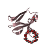
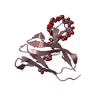
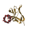
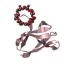
the glycogen-binding domain of the AMP-activated protein kinase
Components
5'-AMP-activated protein kinase, beta-1 subunit
Keywords
SUGAR BINDING PROTEIN / beta sandwich
Function / homology
Function and homology information
AMPK-induced ERAD and lysosome mediated degradation of PD-L1(CD274) / Energy dependent regulation of mTOR by LKB1-AMPK / Regulation of TP53 Activity through Phosphorylation / Macroautophagy / TP53 Regulates Metabolic Genes / nail development / nucleotide-activated protein kinase complex / cellular response to nutrient levels / fatty acid biosynthetic process / positive regulation of cold-induced thermogenesis ...AMPK-induced ERAD and lysosome mediated degradation of PD-L1(CD274) / Energy dependent regulation of mTOR by LKB1-AMPK / Regulation of TP53 Activity through Phosphorylation / Macroautophagy / TP53 Regulates Metabolic Genes / nail development / nucleotide-activated protein kinase complex / cellular response to nutrient levels / fatty acid biosynthetic process / positive regulation of cold-induced thermogenesis / protein kinase binding / signal transduction / protein-containing complex / nucleus / cytosol / cytoplasm Similarity search - Function
Association with the SNF1 complex (ASC) domain / ASC domain superfamily / : / 5'-AMP-activated protein kinase beta subunit, interaction domain / 5'-AMP-activated protein kinase beta subunit, interation domain / AMP-activated protein kinase, glycogen-binding domain / Glycogen recognition site of AMP-activated protein kinase / Immunoglobulin E-set / Immunoglobulin-like fold / Immunoglobulins ...Association with the SNF1 complex (ASC) domain / ASC domain superfamily / : / 5'-AMP-activated protein kinase beta subunit, interaction domain / 5'-AMP-activated protein kinase beta subunit, interation domain / AMP-activated protein kinase, glycogen-binding domain / Glycogen recognition site of AMP-activated protein kinase / Immunoglobulin E-set / Immunoglobulin-like fold / Immunoglobulins / Immunoglobulin-like / Sandwich / Mainly Beta Similarity search - Domain/homology
In the structure databanks used in Yorodumi, some data are registered as the other names, "COVID-19 virus" and "2019-nCoV". Here are the details of the virus and the list of structure data.
Jan 31, 2019. EMDB accession codes are about to change! (news from PDBe EMDB page)
EMDB accession codes are about to change! (news from PDBe EMDB page)
The allocation of 4 digits for EMDB accession codes will soon come to an end. Whilst these codes will remain in use, new EMDB accession codes will include an additional digit and will expand incrementally as the available range of codes is exhausted. The current 4-digit format prefixed with “EMD-” (i.e. EMD-XXXX) will advance to a 5-digit format (i.e. EMD-XXXXX), and so on. It is currently estimated that the 4-digit codes will be depleted around Spring 2019, at which point the 5-digit format will come into force.
The EM Navigator/Yorodumi systems omit the EMD- prefix.
Related info.:Q: What is EMD? / ID/Accession-code notation in Yorodumi/EM Navigator
Yorodumi is a browser for structure data from EMDB, PDB, SASBDB, etc.
This page is also the successor to EM Navigator detail page, and also detail information page/front-end page for Omokage search.
The word "yorodu" (or yorozu) is an old Japanese word meaning "ten thousand". "mi" (miru) is to see.
Related info.:EMDB / PDB / SASBDB / Comparison of 3 databanks / Yorodumi Search / Aug 31, 2016. New EM Navigator & Yorodumi / Yorodumi Papers / Jmol/JSmol / Function and homology information / Changes in new EM Navigator and Yorodumi
 Movie
Movie Controller


 Open data
 Basic information
 Components
 Keywords
 Function and homology information
 X-RAY DIFFRACTION /
  Authors
 Citation
 Structure visualization
 Downloads & links
 Other downloads


 PDBj
PDBj
 Assembly
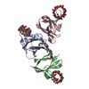


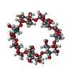

 Mass: 18.015 Da / Num. of mol.: 307 / Source method: isolated from a natural source / Formula: H2O
 Sample preparation
 / Beamline: 14-ID-B / Wavelength: 0.9791 Å
 Processing