 Movie
Movie Controller

[English] 日本語
 Yorodumi
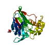
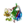
- PDB-3dzp: Thaumatin by LB nanotemplate method after high X-Ray dose on ESRF... -
+ Open data

- Basic information
| Entry | Database: PDB / ID: 3dzp | ||||||
|---|---|---|---|---|---|---|---|
| Title | Thaumatin by LB nanotemplate method after high X-Ray dose on ESRF ID29 beamline | ||||||
 Components Components | Thaumatin-1 | ||||||
 Keywords Keywords | PLANT PROTEIN | ||||||
| Function / homology |  Function and homology information Function and homology information | ||||||
| Biological species |  Thaumatococcus daniellii (katemfe) Thaumatococcus daniellii (katemfe) | ||||||
| Method |  X-RAY DIFFRACTION / SYNCHROTRON / MOLECULAR REPLACEMENT / Resolution: 1.51 Å X-RAY DIFFRACTION / SYNCHROTRON / MOLECULAR REPLACEMENT / Resolution: 1.51 Å | ||||||
 Authors Authors | Tripathi, S. / Pechkova, E. / Nicolini, C. | ||||||
 Citation Citation | Journal: To be Published Title: Radiation damage in protein structural characterization by Synchrotron Radiation: State of the art and Nanotechnology-based perspective Authors: Pechkova, E. / Tripathi, S. / Nicolini, C. | ||||||
| History |
|
- Structure visualization
| Structure viewer | Molecule: MolmilJmol/JSmol |
|---|
- Downloads & links
-Download
| PDBx/mmCIF format | 3dzp.cif.gz | 110.1 KB | Display | PDBx/mmCIF format |
|---|---|---|---|---|
| PDB format | pdb3dzp.ent.gz | 85.2 KB | Display | PDB format |
| PDBx/mmJSON format | 3dzp.json.gz | Tree view | PDBx/mmJSON format | |
| Others |  Other downloads Other downloads |
-Validation report
| Arichive directory | https://data.pdbj.org/pub/pdb/validation_reports/dz/3dzpftp://data.pdbj.org/pub/pdb/validation_reports/dz/3dzp | HTTPS FTP |
|---|
-Related structure data
| Related structure data |  3dnzC  3do0C  3do1C  3do2C  3dvqC  3dvrC 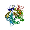 3dvsC  3dw1C 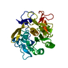 3dw3C  3dweC 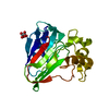 3dznC 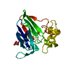 3dzrC 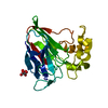 3e0aC 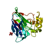 1rqwS C: citing same article ( S: Starting model for refinement |
|---|---|
| Similar structure data |


-Links
 PDBj
PDBj

- Assembly
| Deposited unit | 
| ||||||||
|---|---|---|---|---|---|---|---|---|---|
| 1 |
| ||||||||
| Unit cell |
| ||||||||
| Components on special symmetry positions |
|
-Components
| #1: Protein | Mass: 22243.119 Da / Num. of mol.: 1 / Source method: isolated from a natural source / Source: (natural) Thaumatococcus daniellii (katemfe) / References: UniProt: P02883 |
|---|---|
| #2: Chemical | ChemComp-TLA /   Mass: 150.087 Da / Num. of mol.: 1 / Source method: obtained synthetically / Formula: C4H6O6 Mass: 150.087 Da / Num. of mol.: 1 / Source method: obtained synthetically / Formula: C4H6O6 |
| #3: Water | ChemComp-HOH /  Mass: 18.015 Da / Num. of mol.: 398 / Source method: isolated from a natural source / Formula: H2O Mass: 18.015 Da / Num. of mol.: 398 / Source method: isolated from a natural source / Formula: H2O |
| Has protein modification | Y |
-Experimental details
-Experiment
| Experiment | Method: X-RAY DIFFRACTION / Number of used crystals: 1 |
|---|
- Sample preparation
| Crystal | Density Matthews: 2.82 Å3/Da / Density % sol: 56.35 % / Mosaicity: 0.45 ° |
|---|---|
| Crystal grow | Temperature: 293 K / Method: vapor diffusion, hanging drop / pH: 6.5 Details: 100mM ADA, 1M Na/K-tartrate, pH 6.5, VAPOR DIFFUSION, HANGING DROP, temperature 293K |
-Data collection
| Diffraction | Mean temperature: 110 K |
|---|---|
| Diffraction source | Source: SYNCHROTRON / Site: ESRF  / Beamline: ID29 / Wavelength: 0.9787 Å / Beamline: ID29 / Wavelength: 0.9787 Å |
| Detector | Type: ADSC QUANTUM 315 / Detector: CCD / Date: Apr 29, 2007 / Details: toroidal mirror |
| Radiation | Monochromator: Si 311 / Protocol: SINGLE WAVELENGTH / Monochromatic (M) / Laue (L): M / Scattering type: x-ray |
| Radiation wavelength | Wavelength: 0.9787 Å / Relative weight: 1 |
| Reflection | Resolution: 1.5→53.936 Å / Num. obs: 41813 / % possible obs: 100 % / Redundancy: 7.5 % / Rmerge(I) obs: 0.067 / Rsym value: 0.067 |
| Reflection shell | Resolution: 1.5→1.58 Å / Redundancy: 7.6 % / Rmerge(I) obs: 0.502 / Mean I/σ(I) obs: 1.5 / Num. measured all: 45476 / Num. unique all: 5964 / Rsym value: 0.502 / % possible all: 100 |
- Processing
| Software |
| ||||||||||||||||||||||||||||||||||||||||||||||||||||||||||||||||||||||||||||||||||||||||||
|---|---|---|---|---|---|---|---|---|---|---|---|---|---|---|---|---|---|---|---|---|---|---|---|---|---|---|---|---|---|---|---|---|---|---|---|---|---|---|---|---|---|---|---|---|---|---|---|---|---|---|---|---|---|---|---|---|---|---|---|---|---|---|---|---|---|---|---|---|---|---|---|---|---|---|---|---|---|---|---|---|---|---|---|---|---|---|---|---|---|---|---|
| Refinement | Method to determine structure: MOLECULAR REPLACEMENT Starting model: 1RQW Resolution: 1.51→45.79 Å / Cor.coef. Fo:Fc: 0.969 / Cor.coef. Fo:Fc free: 0.958 / Occupancy max: 1 / Occupancy min: 0.11 / FOM work R set: 0.912 / SU B: 2.23 / SU ML: 0.038 / Cross valid method: THROUGHOUT / σ(F): 0 / ESU R: 0.075 / ESU R Free: 0.064 / Stereochemistry target values: MAXIMUM LIKELIHOOD / Details: HYDROGENS HAVE BEEN ADDED IN THE RIDING POSITIONS
| ||||||||||||||||||||||||||||||||||||||||||||||||||||||||||||||||||||||||||||||||||||||||||
| Solvent computation | Ion probe radii: 0.8 Å / Shrinkage radii: 0.8 Å / VDW probe radii: 1.2 Å / Solvent model: MASK | ||||||||||||||||||||||||||||||||||||||||||||||||||||||||||||||||||||||||||||||||||||||||||
| Displacement parameters | Biso max: 129.94 Å2 / Biso mean: 23.626 Å2 / Biso min: 8.6 Å2
| ||||||||||||||||||||||||||||||||||||||||||||||||||||||||||||||||||||||||||||||||||||||||||
| Refinement step | Cycle: LAST / Resolution: 1.51→45.79 Å
| ||||||||||||||||||||||||||||||||||||||||||||||||||||||||||||||||||||||||||||||||||||||||||
| Refine LS restraints |
| ||||||||||||||||||||||||||||||||||||||||||||||||||||||||||||||||||||||||||||||||||||||||||
| LS refinement shell | Resolution: 1.51→1.549 Å / Total num. of bins used: 20
|
