 Movie
Movie Controller


+ Open data

- Basic information
| Entry | Database: PDB / ID: 3dxf | ||||||
|---|---|---|---|---|---|---|---|
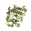
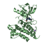
| Title | Crystal structure of the HSCARG R37A mutant | ||||||
 Components Components | NmrA-like family domain-containing protein 1 | ||||||
 Keywords Keywords | NADPH-BINDING PROTEIN / Rossmann fold | ||||||
| Function / homology |  Function and homology information Function and homology informationUrea cycle / perinuclear region of cytoplasm / nucleoplasm / identical protein binding / nucleus / cytosol Similarity search - Function | ||||||
| Biological species |  Homo sapiens (human) Homo sapiens (human) | ||||||
| Method |  X-RAY DIFFRACTION / SYNCHROTRON / MOLECULAR REPLACEMENT / Resolution: 2.2 Å X-RAY DIFFRACTION / SYNCHROTRON / MOLECULAR REPLACEMENT / Resolution: 2.2 Å | ||||||
 Authors Authors | Li, Y. / Meng, G. / Dai, X. / Luo, M. / Zheng, X. | ||||||
 Citation Citation | Journal: J.Mol.Biol. / Year: 2009 Title: NADPH is an allosteric regulator of HSCARG Authors: Dai, X. / Li, Y. / Meng, G. / Yao, S. / Zhao, Y. / Yu, Q. / Zhang, J. / Luo, M. / Zheng, X. | ||||||
| History |
|
- Structure visualization
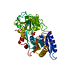
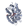
| Structure viewer | Molecule: MolmilJmol/JSmol |
|---|
- Downloads & links
-Download
| PDBx/mmCIF format | 3dxf.cif.gz | 116.9 KB | Display | PDBx/mmCIF format |
|---|---|---|---|---|
| PDB format | pdb3dxf.ent.gz | 91.8 KB | Display | PDB format |
| PDBx/mmJSON format | 3dxf.json.gz | Tree view | PDBx/mmJSON format | |
| Others |  Other downloads Other downloads |
-Validation report
| Arichive directory | https://data.pdbj.org/pub/pdb/validation_reports/dx/3dxfftp://data.pdbj.org/pub/pdb/validation_reports/dx/3dxf | HTTPS FTP |
|---|
-Related structure data
| Related structure data |  3e5mC 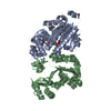 2exxS S: Starting model for refinement C: citing same article ( |
|---|---|
| Similar structure data |


-Links
 PDBj
PDBj
- Assembly
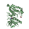
| Deposited unit | 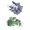
| ||||||||
|---|---|---|---|---|---|---|---|---|---|
| 1 | 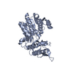
| ||||||||
| 2 | 
| ||||||||
| Unit cell |
|
-Components
| #1: Protein | Mass: 33303.258 Da / Num. of mol.: 2 / Mutation: R37A Source method: isolated from a genetically manipulated source Source: (gene. exp.) Homo sapiens (human) / Plasmid: pET28a / Production host:  |
|---|
-Experimental details
-Experiment
| Experiment | Method: X-RAY DIFFRACTION / Number of used crystals: 1 |
|---|
- Sample preparation
| Crystal | Density Matthews: 2.46 Å3/Da / Density % sol: 50.07 % |
|---|---|
| Crystal grow | Temperature: 277 K / Method: vapor diffusion, hanging drop / pH: 7.2 Details: 1.96M ammonium sulfate, 0.1M sodium Hepes pH 7.2, 2% MPD, 20mM potassium chloride, 5mM Phenol, VAPOR DIFFUSION, HANGING DROP, temperature 277K |
-Data collection
| Diffraction | Mean temperature: 100 K |
|---|---|
| Diffraction source | Source: SYNCHROTRON / Site: BSRF  / Beamline: 3W1A / Wavelength: 0.97928 Å / Beamline: 3W1A / Wavelength: 0.97928 Å |
| Detector | Type: MAR scanner 345 mm plate / Detector: IMAGE PLATE / Date: Mar 17, 2008 |
| Radiation | Protocol: SINGLE WAVELENGTH / Monochromatic (M) / Laue (L): M / Scattering type: x-ray |
| Radiation wavelength | Wavelength: 0.97928 Å / Relative weight: 1 |
| Reflection | Resolution: 2.2→26.7 Å / Num. obs: 32788 / % possible obs: 99.8 % / Observed criterion σ(F): 2 / Observed criterion σ(I): 2 / Redundancy: 5.3 % / Rmerge(I) obs: 0.093 / Net I/σ(I): 16.8 |
| Reflection shell | Resolution: 2.2→2.32 Å / Redundancy: 5.2 % / Rmerge(I) obs: 0.212 / Mean I/σ(I) obs: 6.9 / Num. unique all: 24658 / % possible all: 99.7 |
- Processing
| Software |
| ||||||||||||||||||||||||||||||||||||||||||||||||||||||||||||||||||||||||||||||||||||||||||||||||||||||||||||||||||||||||||||||||||||||||||||||||||||||||||||||||||||||||||
|---|---|---|---|---|---|---|---|---|---|---|---|---|---|---|---|---|---|---|---|---|---|---|---|---|---|---|---|---|---|---|---|---|---|---|---|---|---|---|---|---|---|---|---|---|---|---|---|---|---|---|---|---|---|---|---|---|---|---|---|---|---|---|---|---|---|---|---|---|---|---|---|---|---|---|---|---|---|---|---|---|---|---|---|---|---|---|---|---|---|---|---|---|---|---|---|---|---|---|---|---|---|---|---|---|---|---|---|---|---|---|---|---|---|---|---|---|---|---|---|---|---|---|---|---|---|---|---|---|---|---|---|---|---|---|---|---|---|---|---|---|---|---|---|---|---|---|---|---|---|---|---|---|---|---|---|---|---|---|---|---|---|---|---|---|---|---|---|---|---|---|---|
| Refinement | Method to determine structure: MOLECULAR REPLACEMENT Starting model: PDB ENTRY 2EXX Resolution: 2.2→26 Å / Cor.coef. Fo:Fc: 0.873 / Cor.coef. Fo:Fc free: 0.833 / SU B: 7.102 / SU ML: 0.186 / Cross valid method: THROUGHOUT / ESU R: 0.314 / ESU R Free: 0.24 / Stereochemistry target values: MAXIMUM LIKELIHOOD / Details: HYDROGENS HAVE BEEN ADDED IN THE RIDING POSITIONS
| ||||||||||||||||||||||||||||||||||||||||||||||||||||||||||||||||||||||||||||||||||||||||||||||||||||||||||||||||||||||||||||||||||||||||||||||||||||||||||||||||||||||||||
| Solvent computation | Ion probe radii: 0.8 Å / Shrinkage radii: 0.8 Å / VDW probe radii: 1.2 Å / Solvent model: MASK | ||||||||||||||||||||||||||||||||||||||||||||||||||||||||||||||||||||||||||||||||||||||||||||||||||||||||||||||||||||||||||||||||||||||||||||||||||||||||||||||||||||||||||
| Displacement parameters | Biso mean: 17.598 Å2
| ||||||||||||||||||||||||||||||||||||||||||||||||||||||||||||||||||||||||||||||||||||||||||||||||||||||||||||||||||||||||||||||||||||||||||||||||||||||||||||||||||||||||||
| Refinement step | Cycle: LAST / Resolution: 2.2→26 Å
| ||||||||||||||||||||||||||||||||||||||||||||||||||||||||||||||||||||||||||||||||||||||||||||||||||||||||||||||||||||||||||||||||||||||||||||||||||||||||||||||||||||||||||
| Refine LS restraints |
| ||||||||||||||||||||||||||||||||||||||||||||||||||||||||||||||||||||||||||||||||||||||||||||||||||||||||||||||||||||||||||||||||||||||||||||||||||||||||||||||||||||||||||
| LS refinement shell | Resolution: 2.2→2.257 Å / Total num. of bins used: 20
|
