 Movie
Movie Controller

[English] 日本語
 Yorodumi
- PDB-3dry: X-ray crystal structure of human KCTD5 protein crystallized in lo... -
+ Open data

- Basic information
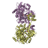
| Entry | Database: PDB / ID: 3dry | ||||||
|---|---|---|---|---|---|---|---|
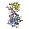
| Title | X-ray crystal structure of human KCTD5 protein crystallized in low-salt buffer | ||||||
 Components Components | BTB/POZ domain-containing protein KCTD5 | ||||||
 Keywords Keywords | UNKNOWN FUNCTION / KCTD5 / BTB/POZ / Golgi / GRASP55 / potassium channel DOMAIN T1 / PENTAMERIC ASSEMBLY / Host-virus interaction / Nucleus | ||||||
| Function / homology |  Function and homology information Function and homology informationCul3-RING ubiquitin ligase complex / cullin family protein binding / protein homooligomerization / proteasome-mediated ubiquitin-dependent protein catabolic process / protein-containing complex binding / identical protein binding / nucleus / cytoplasm / cytosol Similarity search - Function | ||||||
| Biological species |  Homo sapiens (human) Homo sapiens (human) | ||||||
| Method |  X-RAY DIFFRACTION / SYNCHROTRON / MOLECULAR REPLACEMENT / Resolution: 3.3 Å X-RAY DIFFRACTION / SYNCHROTRON / MOLECULAR REPLACEMENT / Resolution: 3.3 Å | ||||||
 Authors Authors | Tereshko, V. / Dementieva, I. / Goldstein, S.A.N. | ||||||
 Citation Citation | Journal: J.Mol.Biol. / Year: 2009 Title: Pentameric assembly of potassium channel tetramerization domain-containing protein 5. Authors: Dementieva, I.S. / Tereshko, V. / McCrossan, Z.A. / Solomaha, E. / Araki, D. / Xu, C. / Grigorieff, N. / Goldstein, S.A. | ||||||
| History |
|
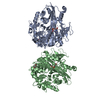
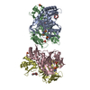
- Structure visualization
| Structure viewer | Molecule: MolmilJmol/JSmol |
|---|
- Downloads & links
-Download
| PDBx/mmCIF format | 3dry.cif.gz | 174.2 KB | Display | PDBx/mmCIF format |
|---|---|---|---|---|
| PDB format | pdb3dry.ent.gz | 139.2 KB | Display | PDB format |
| PDBx/mmJSON format | 3dry.json.gz | Tree view | PDBx/mmJSON format | |
| Others |  Other downloads Other downloads |
-Validation report
| Arichive directory | https://data.pdbj.org/pub/pdb/validation_reports/dr/3dryftp://data.pdbj.org/pub/pdb/validation_reports/dr/3dry | HTTPS FTP |
|---|
-Related structure data
| Related structure data |  3drxSC 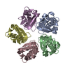 3drzC S: Starting model for refinement C: citing same article ( |
|---|---|
| Similar structure data |
-Links
 PDBj
PDBj


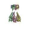
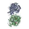
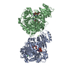
- Assembly
| Deposited unit | 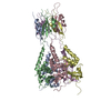
| ||||||||||||||||||||||||||||||||||||||||||||||||||||||||||||||||||||||||||||||||||||||||||||||||||||||||||||||||||||||||||||||||||||||||||||||||||||||||||||||||||||||||||||||||||||||||||||||||||||||||
|---|---|---|---|---|---|---|---|---|---|---|---|---|---|---|---|---|---|---|---|---|---|---|---|---|---|---|---|---|---|---|---|---|---|---|---|---|---|---|---|---|---|---|---|---|---|---|---|---|---|---|---|---|---|---|---|---|---|---|---|---|---|---|---|---|---|---|---|---|---|---|---|---|---|---|---|---|---|---|---|---|---|---|---|---|---|---|---|---|---|---|---|---|---|---|---|---|---|---|---|---|---|---|---|---|---|---|---|---|---|---|---|---|---|---|---|---|---|---|---|---|---|---|---|---|---|---|---|---|---|---|---|---|---|---|---|---|---|---|---|---|---|---|---|---|---|---|---|---|---|---|---|---|---|---|---|---|---|---|---|---|---|---|---|---|---|---|---|---|---|---|---|---|---|---|---|---|---|---|---|---|---|---|---|---|---|---|---|---|---|---|---|---|---|---|---|---|---|---|---|---|---|
| 1 |
| ||||||||||||||||||||||||||||||||||||||||||||||||||||||||||||||||||||||||||||||||||||||||||||||||||||||||||||||||||||||||||||||||||||||||||||||||||||||||||||||||||||||||||||||||||||||||||||||||||||||||
| Unit cell |
| ||||||||||||||||||||||||||||||||||||||||||||||||||||||||||||||||||||||||||||||||||||||||||||||||||||||||||||||||||||||||||||||||||||||||||||||||||||||||||||||||||||||||||||||||||||||||||||||||||||||||
| Noncrystallographic symmetry (NCS) | NCS domain:
NCS domain segments: Refine code: 4
|