[English] 日本語
Yorodumi
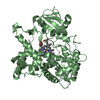
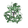
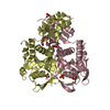
- PDB-1o57: CRYSTAL STRUCTURE OF THE PURINE OPERON REPRESSOR OF BACILLUS SUBTILIS -

+
Open data


ID or keywords:

Loading...

-
Basic information

Entry
Database: PDB / ID: 1o57
TitleCRYSTAL STRUCTURE OF THE PURINE OPERON REPRESSOR OF BACILLUS SUBTILIS
ComponentsPUR OPERON REPRESSOR
KeywordsDNA BINDING PROTEIN / PURINE OPERON REPRESSOR / HELIX-TURN-HELIX DOMAIN / PHOSPHORIBOSYLTRANSEFERASES / DOMAIN RECOMBINATION / DNA BINDING / TRANSCRIPTION REGULATION
Function / homology
Function and homology information


negative regulation of purine nucleobase metabolic process / negative regulation of DNA-templated transcription / DNA binding
Similarity search - Function
Pur operon repressor / Bacterial purine repressor, N-terminal / Bacterial purine repressor, N-terminal / : / Rossmann fold - #2020 / Phosphoribosyl transferase domain / Phosphoribosyltransferase-like / Phosphoribosyltransferase domain / Winged helix-like DNA-binding domain superfamily/Winged helix DNA-binding domain / Arc Repressor Mutant, subunit A ...Pur operon repressor / Bacterial purine repressor, N-terminal / Bacterial purine repressor, N-terminal / : / Rossmann fold - #2020 / Phosphoribosyl transferase domain / Phosphoribosyltransferase-like / Phosphoribosyltransferase domain / Winged helix-like DNA-binding domain superfamily/Winged helix DNA-binding domain / Arc Repressor Mutant, subunit A / Winged helix DNA-binding domain superfamily / Winged helix-like DNA-binding domain superfamily / Rossmann fold / Orthogonal Bundle / 3-Layer(aba) Sandwich / Mainly Alpha / Alpha Beta
Similarity search - Domain/homology
Pur operon repressor
Similarity search - Component
Biological speciesBacillus subtilis (bacteria)
MethodX-RAY DIFFRACTION / SYNCHROTRON / MAD / Resolution: 2.2 Å
AuthorsSinha, S.C. / Krahn, J. / Shin, B.S. / Tomchick, D.R. / Zalkin, H. / Smith, J.L.
CitationJournal: J.Bacteriol. / Year: 2003
Title: The Purine Repressor of Bacillus Subtilis: A Novel Combination of Domains Adapted for Transcription Regulation
Authors: Sinha, S.C. / Krahn, J. / Shin, B.S. / Tomchick, D.R. / Zalkin, H. / Smith, J.L.
History
DepositionApr 20, 2003Deposition site: RCSB / Processing site: RCSB
SupersessionAug 26, 2003ID: 1P41
Revision 1.0Aug 26, 2003Provider: repository / Type: Initial release
Revision 1.1Apr 26, 2008Group: Version format compliance
Revision 1.2Jul 13, 2011Group: Version format compliance
Revision 1.3Dec 27, 2023Group: Data collection / Database references / Derived calculations
Category: chem_comp_atom / chem_comp_bond ...chem_comp_atom / chem_comp_bond / database_2 / struct_ref_seq_dif / struct_site
Item: _database_2.pdbx_DOI / _database_2.pdbx_database_accession ..._database_2.pdbx_DOI / _database_2.pdbx_database_accession / _struct_ref_seq_dif.details / _struct_site.pdbx_auth_asym_id / _struct_site.pdbx_auth_comp_id / _struct_site.pdbx_auth_seq_id

-
Structure visualization

Structure viewerMolecule:
MolmilJmol/JSmol

Downloads & links

-
Assembly

Deposited unit
A: PUR OPERON REPRESSOR
B: PUR OPERON REPRESSOR
C: PUR OPERON REPRESSOR
D: PUR OPERON REPRESSOR
hetero molecules


Theoretical massNumber of molelcules
Total (without water)131,25420
Polymers128,3594
Non-polymers2,89516
Water10,233568
1
A: PUR OPERON REPRESSOR
B: PUR OPERON REPRESSOR
hetero molecules


Theoretical massNumber of molelcules
Total (without water)65,69310
Polymers64,1802
Non-polymers1,5148
Water362
TypeNameSymmetry operationNumber
identity operation1_555x,y,z1
Buried area6610 Å2
ΔGint-63 kcal/mol
Surface area23010 Å2
MethodPISA
2
C: PUR OPERON REPRESSOR
D: PUR OPERON REPRESSOR
hetero molecules


Theoretical massNumber of molelcules
Total (without water)65,56110
Polymers64,1802
Non-polymers1,3818
Water362
TypeNameSymmetry operationNumber
identity operation1_555x,y,z1
Buried area6500 Å2
ΔGint-76 kcal/mol
Surface area22630 Å2
MethodPISA
Unit cell
Length a, b, c (Å)65.077, 72.156, 82.967
Angle α, β, γ (deg.)84.75, 84.03, 67.47
Int Tables number1
Space group name H-MP1

-
Components

-
Protein , 1 types, 4 molecules ABCD

#1: Protein
PUR OPERON REPRESSOR


Mass: 32089.771 Da / Num. of mol.: 4
Source method: isolated from a genetically manipulated source
Source: (gene. exp.) Bacillus subtilis (bacteria) / Gene: PURR / Production host: Escherichia coli (E. coli) / Strain (production host): B834(DE3) / References: UniProt: P37551

-
Non-polymers , 7 types, 584 molecules

#2: Chemical
ChemComp-SO4 / SULFATE ION


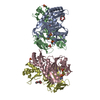
Mass: 96.063 Da / Num. of mol.: 8 / Source method: obtained synthetically / Formula: SO4
#3: Chemical ChemComp-EPE / 4-(2-HYDROXYETHYL)-1-PIPERAZINE ETHANESULFONIC ACID / HEPES


Mass: 238.305 Da / Num. of mol.: 2 / Source method: obtained synthetically / Formula: C8H18N2O4S / Comment: pH buffer*YM
#4: Chemical ChemComp-P6G / HEXAETHYLENE GLYCOL / POLYETHYLENE GLYCOL PEG400


Mass: 282.331 Da / Num. of mol.: 2 / Source method: obtained synthetically / Formula: C12H26O7 / Comment: precipitant*YM
#5: Chemical ChemComp-2PE / NONAETHYLENE GLYCOL


Mass: 414.488 Da / Num. of mol.: 1 / Source method: obtained synthetically / Formula: C18H38O10 / Comment: precipitant*YM
#6: Chemical ChemComp-PG4 / TETRAETHYLENE GLYCOL


Mass: 194.226 Da / Num. of mol.: 1 / Source method: obtained synthetically / Formula: C8H18O5 / Comment: precipitant*YM
#7: Chemical ChemComp-1PE / PENTAETHYLENE GLYCOL / PEG400


Mass: 238.278 Da / Num. of mol.: 2 / Source method: obtained synthetically / Formula: C10H22O6 / Comment: precipitant*YM
#8: Water ChemComp-HOH / water


Mass: 18.015 Da / Num. of mol.: 568 / Source method: isolated from a natural source / Formula: H2O

-
Experimental details

-
Experiment

ExperimentMethod: X-RAY DIFFRACTION / Number of used crystals: 1

-
Sample preparation

CrystalDensity Matthews: 2.8 Å3/Da / Density % sol: 55.6 %
Crystal growTemperature: 293 K / Method: vapor diffusion, hanging drop / pH: 7
Details: 10mg/mL PurR, 83 mM HEPES, pH 7.0, 5% PEG8000, 450 mM Lithium sulfate, 50 mM ammonium sulfate, 300 mM NaCl, 5mM DTT, VAPOR DIFFUSION, HANGING DROP, temperature 293K, pH 7.00
Crystal grow
*PLUS
Temperature: 20 ℃ / pH: 8 / Method: vapor diffusion, hanging drop
Components of the solutions
*PLUS
IDConc.Common nameCrystal-IDSol-IDDetailsChemical formula
110 mg/mlprotein1drop
210 mMHEPES1droppH8.0
350 mMammonium sulfate1drop
4300 mM1dropNaCl
55 %PEG80001reservoir
666.7 mMHEPES1reservoirpH7.0
7167 mM1reservoirLi2SO4

-
Data collection

Diffraction
IDMean temperature (K)Crystal-ID
11001
21
Diffraction sourceSource: SYNCHROTRON / Site: ESRF / Beamline: ID14-1 / Wavelength: 0.9790, 0.9788, 1.5418
Detector
TypeIDDetectorDate
MARRESEARCH1IMAGE PLATESep 1, 1997
RIGAKU RAXIS IIC2IMAGE PLATESep 1, 1997
RadiationProtocol: MAD / Monochromatic (M) / Laue (L): M / Scattering type: x-ray
Radiation wavelength
IDWavelength (Å)Relative weight
10.9791
20.97881
31.54181
ReflectionResolution: 2→20 Å / Num. obs: 66722 / % possible obs: 97.6 % / Observed criterion σ(I): 2 / Redundancy: 4.3 % / Biso Wilson estimate: 45.3 Å2 / Rsym value: 0.073 / Net I/σ(I): 11.1
Reflection shellResolution: 2→2.28 Å / Redundancy: 1 % / Mean I/σ(I) obs: 3.6 / Rsym value: 0.194 / % possible all: 96.6
Reflection
*PLUS
Highest resolution: 2.2 Å / Lowest resolution: 30 Å / Redundancy: 4.3 % / Rmerge(I) obs: 0.073
Reflection shell
*PLUS
Highest resolution: 2.2 Å / % possible obs: 96.6 % / Rmerge(I) obs: 0.194 / Mean I/σ(I) obs: 3.6

-
Processing

Software
NameVersionClassification
DENZOdata reduction
SCALEPACKdata scaling
MLPHAREphasing
CNS1.1refinement
RefinementMethod to determine structure: MAD / Resolution: 2.2→20 Å / Rfactor Rfree error: 0.004 / Data cutoff high absF: 1384607.27 / Data cutoff low absF: 0 / Isotropic thermal model: RESTRAINED / Cross valid method: THROUGHOUT / σ(F): 0 / Stereochemistry target values: ENGH & HUBER
Details: NCS RESTRAINTS WERE USED IN ALL BUT THE FINAL CYCLES OF REFINEMENT
RfactorNum. reflection% reflectionSelection details
Rfree0.237 3335 5 %RANDOM
Rwork0.188 ---
obs0.1 66365 94.4 %-
Solvent computationSolvent model: FLAT MODEL / Bsol: 52.05 Å2 / ksol: 0.33 e/Å3
Displacement parametersBiso mean: 52.7 Å2
Baniso -1Baniso -2Baniso -3
1-1.35 Å22.47 Å2-0.65 Å2
2--0.88 Å20.16 Å2
3----2.23 Å2
Refine analyze
FreeObs
Luzzati coordinate error0.32 Å0.24 Å
Luzzati d res low-5 Å
Luzzati sigma a0.3 Å0.21 Å
Refinement stepCycle: LAST / Resolution: 2.2→20 Å
ProteinNucleic acidLigandSolventTotal
Num. atoms8383 0 181 568 9132
Refine LS restraints
Refine-IDTypeDev idealDev ideal target
X-RAY DIFFRACTIONc_bond_d0.01
X-RAY DIFFRACTIONc_bond_d_na
X-RAY DIFFRACTIONc_bond_d_prot
X-RAY DIFFRACTIONc_angle_d
X-RAY DIFFRACTIONc_angle_d_na
X-RAY DIFFRACTIONc_angle_d_prot
X-RAY DIFFRACTIONc_angle_deg1.5
X-RAY DIFFRACTIONc_angle_deg_na
X-RAY DIFFRACTIONc_angle_deg_prot
X-RAY DIFFRACTIONc_dihedral_angle_d23.5
X-RAY DIFFRACTIONc_dihedral_angle_d_na
X-RAY DIFFRACTIONc_dihedral_angle_d_prot
X-RAY DIFFRACTIONc_improper_angle_d0.93
X-RAY DIFFRACTIONc_improper_angle_d_na
X-RAY DIFFRACTIONc_improper_angle_d_prot
X-RAY DIFFRACTIONc_mcbond_it4.394.5
X-RAY DIFFRACTIONc_mcangle_it65
X-RAY DIFFRACTIONc_scbond_it7.86
X-RAY DIFFRACTIONc_scangle_it9.747.5
LS refinement shellResolution: 2.2→2.34 Å / Rfactor Rfree error: 0.014 / Total num. of bins used: 6
RfactorNum. reflection% reflection
Rfree0.308 515 4.9 %
Rwork0.233 9938 -
obs--89.2 %
Xplor file
Refine-IDSerial noParam fileTopol file
X-RAY DIFFRACTION1PROTEIN_REP.PARAMPROTEIN.TOP
X-RAY DIFFRACTION2WATER_REP.PARAMWATER_REP.TOP
X-RAY DIFFRACTION3LIGANDS.PARLIGANDS.TOP
X-RAY DIFFRACTION4ION.PARAMION.TOP
X-RAY DIFFRACTION5
Refinement
*PLUS
Highest resolution: 2.2 Å / Lowest resolution: 20 Å / % reflection Rfree: 4.7 %
Solvent computation
*PLUS
Displacement parameters
*PLUS
Refine LS restraints
*PLUS
Refine-IDTypeDev ideal
X-RAY DIFFRACTIONc_angle_deg1.459
X-RAY DIFFRACTIONc_dihedral_angle_d
X-RAY DIFFRACTIONc_dihedral_angle_deg23.5
X-RAY DIFFRACTIONc_improper_angle_d
X-RAY DIFFRACTIONc_improper_angle_deg0.93

+
About Yorodumi

-
News

-
Feb 9, 2022. New format data for meta-information of EMDB entries

New format data for meta-information of EMDB entries

  • Version 3 of the EMDB header file is now the official format.
  • The previous official version 1.9 will be removed from the archive.

Related info.:EMDB header

External links:wwPDB to switch to version 3 of the EMDB data model

-
Aug 12, 2020. Covid-19 info

Covid-19 info

URL: https://pdbjlvh1.pdbj.org/emnavi/covid19.php

New page: Covid-19 featured information page in EM Navigator.

Related info.:Covid-19 info / Mar 5, 2020. Novel coronavirus structure data

+
Mar 5, 2020. Novel coronavirus structure data

Novel coronavirus structure data

Related info.:Yorodumi Speices / Aug 12, 2020. Covid-19 info

External links:COVID-19 featured content - PDBj / Molecule of the Month (242):Coronavirus Proteases

+
Jan 31, 2019. EMDB accession codes are about to change! (news from PDBe EMDB page)

EMDB accession codes are about to change! (news from PDBe EMDB page)

  • The allocation of 4 digits for EMDB accession codes will soon come to an end. Whilst these codes will remain in use, new EMDB accession codes will include an additional digit and will expand incrementally as the available range of codes is exhausted. The current 4-digit format prefixed with “EMD-” (i.e. EMD-XXXX) will advance to a 5-digit format (i.e. EMD-XXXXX), and so on. It is currently estimated that the 4-digit codes will be depleted around Spring 2019, at which point the 5-digit format will come into force.
  • The EM Navigator/Yorodumi systems omit the EMD- prefix.

Related info.:Q: What is EMD? / ID/Accession-code notation in Yorodumi/EM Navigator

External links:EMDB Accession Codes are Changing Soon! / Contact to PDBj

+
Jul 12, 2017. Major update of PDB

Major update of PDB

  • wwPDB released updated PDB data conforming to the new PDBx/mmCIF dictionary.
  • This is a major update changing the version number from 4 to 5, and with Remediation, in which all the entries are updated.
  • In this update, many items about electron microscopy experimental information are reorganized (e.g. em_software).
  • Now, EM Navigator and Yorodumi are based on the updated data.

External links:wwPDB Remediation / Enriched Model Files Conforming to OneDep Data Standards Now Available in the PDB FTP Archive

-
Yorodumi

Thousand views of thousand structures

  • Yorodumi is a browser for structure data from EMDB, PDB, SASBDB, etc.
  • This page is also the successor to EM Navigator detail page, and also detail information page/front-end page for Omokage search.
  • The word "yorodu" (or yorozu) is an old Japanese word meaning "ten thousand". "mi" (miru) is to see.

Related info.:EMDB / PDB / SASBDB / Comparison of 3 databanks / Yorodumi Search / Aug 31, 2016. New EM Navigator & Yorodumi / Yorodumi Papers / Jmol/JSmol / Function and homology information / Changes in new EM Navigator and Yorodumi

Read more