 Movie
Movie Controller

[English] 日本語
 Yorodumi
- PDB-3dqp: Crystal structure of the oxidoreductase ylbE from Lactococcus lac... -
+ Open data

- Basic information
| Entry | Database: PDB / ID: 3dqp | ||||||
|---|---|---|---|---|---|---|---|
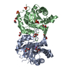
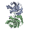
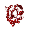
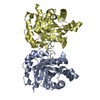
| Title | Crystal structure of the oxidoreductase ylbE from Lactococcus lactis, Northeast Structural Genomics Consortium Target KR121. | ||||||
 Components Components | Oxidoreductase ylbE | ||||||
 Keywords Keywords | OXIDOREDUCTASE / alpha-beta protein. / Structural Genomics / PSI-2 / Protein Structure Initiative / Northeast Structural Genomics Consortium / NESG | ||||||
| Function / homology | NAD(P)H-binding / NAD(P)-binding domain / NAD(P)-binding Rossmann-like Domain / NAD(P)-binding domain superfamily / Rossmann fold / 3-Layer(aba) Sandwich / Alpha Beta / Oxidoreductase Function and homology information Function and homology information | ||||||
| Biological species |  Lactococcus lactis subsp. lactis (lactic acid bacteria) Lactococcus lactis subsp. lactis (lactic acid bacteria) | ||||||
| Method |  X-RAY DIFFRACTION / SYNCHROTRON / SAD / Resolution: 1.4 Å X-RAY DIFFRACTION / SYNCHROTRON / SAD / Resolution: 1.4 Å | ||||||
 Authors Authors | Forouhar, F. / Neely, H. / Seetharaman, J. / Mao, L. / Xiao, R. / Ciccosanti, C. / Foote, E.L. / Lee, D. / Everett, J.K. / Acton, T.B. ...Forouhar, F. / Neely, H. / Seetharaman, J. / Mao, L. / Xiao, R. / Ciccosanti, C. / Foote, E.L. / Lee, D. / Everett, J.K. / Acton, T.B. / Montelione, G.T. / Tong, L. / Hunt, J.F. / Northeast Structural Genomics Consortium (NESG) | ||||||
 Citation Citation | Journal: To be Published Title: Crystal structure of the oxidoreductase ylbE from Lactococcus lactis, Northeast Structural Genomics Consortium Target KR121. Authors: Forouhar, F. / Neely, H. / Seetharaman, J. / Mao, L. / Xiao, R. / Ciccosanti, C. / Foote, E.L. / Lee, D. / Everett, J.K. / B Acton, T. / Montelione, G.T. / Tong, L. / Hunt, J.F. | ||||||
| History |
|
- Structure visualization
| Structure viewer | Molecule: MolmilJmol/JSmol |
|---|
- Downloads & links
-Download
| PDBx/mmCIF format | 3dqp.cif.gz | 65.9 KB | Display | PDBx/mmCIF format |
|---|---|---|---|---|
| PDB format | pdb3dqp.ent.gz | 48 KB | Display | PDB format |
| PDBx/mmJSON format | 3dqp.json.gz | Tree view | PDBx/mmJSON format | |
| Others |  Other downloads Other downloads |
-Validation report
| Arichive directory | https://data.pdbj.org/pub/pdb/validation_reports/dq/3dqpftp://data.pdbj.org/pub/pdb/validation_reports/dq/3dqp | HTTPS FTP |
|---|
-Related structure data
| Similar structure data | |
|---|---|
| Other databases |


-Links
 PDBj
PDBj- Assembly
| Deposited unit | 
| ||||||||
|---|---|---|---|---|---|---|---|---|---|
| 1 | 
| ||||||||
| Unit cell |
| ||||||||
| Details | authors state that the biological unit is possibly dimer. |
-Components
| #1: Protein | Mass: 24523.037 Da / Num. of mol.: 1 / Mutation: I64M Source method: isolated from a genetically manipulated source Source: (gene. exp.) Lactococcus lactis subsp. lactis (lactic acid bacteria)Strain: IL1403 / Gene: ylbE, LL1109, L119, L119013 / Plasmid: pET21 / Production host: |
|---|---|
| #2: Water | ChemComp-HOH /  Mass: 18.015 Da / Num. of mol.: 464 / Source method: isolated from a natural source / Formula: H2O Mass: 18.015 Da / Num. of mol.: 464 / Source method: isolated from a natural source / Formula: H2O |
| Has protein modification | Y |
-Experimental details
-Experiment
| Experiment | Method: X-RAY DIFFRACTION / Number of used crystals: 1 |
|---|
- Sample preparation
| Crystal | Density Matthews: 2.32 Å3/Da / Density % sol: 47 % |
|---|---|
| Crystal grow | Temperature: 291 K / Method: microbatch, under oil / pH: 7.5 Details: Protein solution: 10 mM Tris (pH 7.5), 100 mM sodium chloride, and 5 mM DTT. Reservoir solution: 0.1M HEPES (pH 7.5), 40% PEG 20K, and 0.1M NaMolybdate., microbatch, under oil, temperature 291K |
-Data collection
| Diffraction | Mean temperature: 100 K |
|---|---|
| Diffraction source | Source: SYNCHROTRON / Site: NSLS  / Beamline: X4C / Wavelength: 0.97862 Å / Beamline: X4C / Wavelength: 0.97862 Å |
| Detector | Type: MAR CCD 165 mm / Detector: CCD / Date: Jul 2, 2008 / Details: mirrors |
| Radiation | Monochromator: Si 111 CHANNEL / Protocol: SINGLE WAVELENGTH / Monochromatic (M) / Laue (L): M / Scattering type: x-ray |
| Radiation wavelength | Wavelength: 0.97862 Å / Relative weight: 1 |
| Reflection | Resolution: 1.4→30 Å / Num. all: 87349 / Num. obs: 78964 / % possible obs: 90.4 % / Observed criterion σ(F): 0 / Observed criterion σ(I): 0 / Redundancy: 3.3 % / Biso Wilson estimate: 10.2 Å2 / Rmerge(I) obs: 0.06 / Rsym value: 0.054 / Net I/σ(I): 23.6 |
| Reflection shell | Resolution: 1.4→1.45 Å / Redundancy: 3.4 % / Rmerge(I) obs: 0.234 / Mean I/σ(I) obs: 4.59 / Num. unique all: 8724 / Rsym value: 0.18 / % possible all: 99.7 |
- Processing
| Software |
| |||||||||||||||||||||||||
|---|---|---|---|---|---|---|---|---|---|---|---|---|---|---|---|---|---|---|---|---|---|---|---|---|---|---|
| Refinement | Method to determine structure: SAD / Resolution: 1.4→9.84 Å / Rfactor Rfree error: 0.003 / Data cutoff high absF: 852247.64 / Data cutoff low absF: 0 / Isotropic thermal model: OVERALL / Cross valid method: THROUGHOUT / σ(F): 2 / σ(I): 2 / Stereochemistry target values: Engh & Huber
| |||||||||||||||||||||||||
| Solvent computation | Solvent model: FLAT MODEL / Bsol: 60.3821 Å2 / ksol: 0.45 e/Å3 | |||||||||||||||||||||||||
| Displacement parameters | Biso mean: 15.6 Å2
| |||||||||||||||||||||||||
| Refine analyze |
| |||||||||||||||||||||||||
| Refinement step | Cycle: LAST / Resolution: 1.4→9.84 Å
| |||||||||||||||||||||||||
| Refine LS restraints |
| |||||||||||||||||||||||||
| LS refinement shell | Resolution: 1.4→1.45 Å / Rfactor Rfree error: 0.015 / Total num. of bins used: 10
|
