 Movie
Movie Controller

[English] 日本語
 Yorodumi
- PDB-3dpi: Crystal Structure of NAD+ synthetase from Burkholderia Pseudomallei -
+ Open data

- Basic information
| Entry | Database: PDB / ID: 3dpi | ||||||
|---|---|---|---|---|---|---|---|
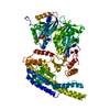
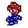
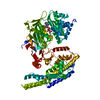
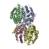
| Title | Crystal Structure of NAD+ synthetase from Burkholderia Pseudomallei | ||||||
 Components Components | NAD+ synthetase | ||||||
 Keywords Keywords | LIGASE / SSGCID / deCODE / Structural Genomics / PSI / Protein Structure Initiative / Seattle Structural Genomics Center for Infectious Disease | ||||||
| Function / homology |  Function and homology information Function and homology informationNAD+ synthase / NAD+ synthase activity / NAD+ synthase (glutamine-hydrolyzing) activity / glutaminase activity / NAD+ biosynthetic process / ATP binding / metal ion binding / cytoplasm Similarity search - Function | ||||||
| Biological species |  Burkholderia pseudomallei (bacteria) Burkholderia pseudomallei (bacteria) | ||||||
| Method |  X-RAY DIFFRACTION / SYNCHROTRON / MOLECULAR REPLACEMENT / molecular replacement / Resolution: 2.2 Å X-RAY DIFFRACTION / SYNCHROTRON / MOLECULAR REPLACEMENT / molecular replacement / Resolution: 2.2 Å | ||||||
 Authors Authors | Seattle Structural Genomics Center for Infectious Disease (SSGCID) | ||||||
 Citation Citation | Journal: To be Published Title: Crystal Structure of NAD+ synthetase from Burkholderia Pseudomallei Authors: Seattle Structural Genomics Center for Infectious Disease (SSGCID) | ||||||
| History |
|
- Structure visualization
| Structure viewer | Molecule: MolmilJmol/JSmol |
|---|
- Downloads & links
-Download
| PDBx/mmCIF format | 3dpi.cif.gz | 110.6 KB | Display | PDBx/mmCIF format |
|---|---|---|---|---|
| PDB format | pdb3dpi.ent.gz | 84.4 KB | Display | PDB format |
| PDBx/mmJSON format | 3dpi.json.gz | Tree view | PDBx/mmJSON format | |
| Others |  Other downloads Other downloads |
-Validation report
| Arichive directory | https://data.pdbj.org/pub/pdb/validation_reports/dp/3dpiftp://data.pdbj.org/pub/pdb/validation_reports/dp/3dpi | HTTPS FTP |
|---|
-Related structure data
| Related structure data | 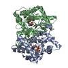 2pz8S S: Starting model for refinement |
|---|---|
| Similar structure data |
-Links
 PDBj
PDBj- Assembly
| Deposited unit | 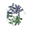
| ||||||||
|---|---|---|---|---|---|---|---|---|---|
| 1 | 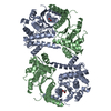
| ||||||||
| 2 |
| ||||||||
| Unit cell |
|
-Components
| #1: Protein | Mass: 31012.156 Da / Num. of mol.: 2 Source method: isolated from a genetically manipulated source Source: (gene. exp.) Burkholderia pseudomallei (bacteria) / Strain: 1710b / Gene: 76819159 / Production host: #2: Chemical | ChemComp-ACT / |   Mass: 59.044 Da / Num. of mol.: 1 / Source method: obtained synthetically / Formula: C2H3O2 Mass: 59.044 Da / Num. of mol.: 1 / Source method: obtained synthetically / Formula: C2H3O2#3: Water | ChemComp-HOH / |  Mass: 18.015 Da / Num. of mol.: 229 / Source method: isolated from a natural source / Formula: H2O Mass: 18.015 Da / Num. of mol.: 229 / Source method: isolated from a natural source / Formula: H2O |
|---|
-Experimental details
-Experiment
| Experiment | Method: X-RAY DIFFRACTION / Number of used crystals: 1 |
|---|
- Sample preparation
| Crystal | Density Matthews: 2.16 Å3/Da / Density % sol: 42.98 % |
|---|---|
| Crystal grow | Temperature: 289 K / Method: vapor diffusion, sitting drop / pH: 5.5 Details: 25% PEG 3350, 0.2 M NaCl, 0.1 M BisTris pH 5.5, Hampton Index screen well F10, Crystal ID 109415F10, VAPOR DIFFUSION, SITTING DROP, temperature 289K |
-Data collection
| Diffraction | Mean temperature: 100 K |
|---|---|
| Diffraction source | Source: SYNCHROTRON / Site: SSRL  / Beamline: BL7-1 / Wavelength: 0.97607 Å / Beamline: BL7-1 / Wavelength: 0.97607 Å |
| Detector | Date: May 27, 2008 |
| Radiation | Protocol: SINGLE WAVELENGTH / Monochromatic (M) / Laue (L): M / Scattering type: x-ray |
| Radiation wavelength | Wavelength: 0.97607 Å / Relative weight: 1 |
| Reflection | Resolution: 2.2→50 Å / Num. obs: 28418 / % possible obs: 99.9 % / Redundancy: 12.6 % / Rmerge(I) obs: 0.086 / Χ2: 1.286 / Net I/σ(I): 15.2 |
| Reflection shell | Resolution: 2.2→2.28 Å / Redundancy: 13 % / Rmerge(I) obs: 0.447 / Mean I/σ(I) obs: 6.1 / Num. unique all: 2778 / Χ2: 1.236 / % possible all: 100 |
-Phasing
| Phasing | Method: molecular replacement |
|---|
- Processing
| Software |
| |||||||||||||||||||||||||||||||||||||||||||||||||||||||||||||||||
|---|---|---|---|---|---|---|---|---|---|---|---|---|---|---|---|---|---|---|---|---|---|---|---|---|---|---|---|---|---|---|---|---|---|---|---|---|---|---|---|---|---|---|---|---|---|---|---|---|---|---|---|---|---|---|---|---|---|---|---|---|---|---|---|---|---|---|
| Refinement | Method to determine structure: MOLECULAR REPLACEMENT Starting model: 2PZ8 Resolution: 2.2→50 Å / Cor.coef. Fo:Fc: 0.937 / Cor.coef. Fo:Fc free: 0.927 / Occupancy max: 1 / Occupancy min: 0.5 / SU B: 6.188 / SU ML: 0.158 / Cross valid method: THROUGHOUT / σ(F): 0 / ESU R: 0.303 / ESU R Free: 0.213 / Stereochemistry target values: MAXIMUM LIKELIHOOD / Details: HYDROGENS HAVE BEEN ADDED IN THE RIDING POSITIONS
| |||||||||||||||||||||||||||||||||||||||||||||||||||||||||||||||||
| Solvent computation | Ion probe radii: 0.8 Å / Shrinkage radii: 0.8 Å / VDW probe radii: 1.2 Å / Solvent model: MASK | |||||||||||||||||||||||||||||||||||||||||||||||||||||||||||||||||
| Displacement parameters | Biso max: 80.44 Å2 / Biso mean: 42.93 Å2 / Biso min: 20 Å2
| |||||||||||||||||||||||||||||||||||||||||||||||||||||||||||||||||
| Refinement step | Cycle: LAST / Resolution: 2.2→50 Å
| |||||||||||||||||||||||||||||||||||||||||||||||||||||||||||||||||
| Refine LS restraints |
| |||||||||||||||||||||||||||||||||||||||||||||||||||||||||||||||||
| LS refinement shell | Resolution: 2.2→2.257 Å / Total num. of bins used: 20
|
